Cytology of the scrotum
Introduction
Most pathology of the scrotum involves the scrotal skin. Lesions can be due to a large variety of etiologies including trauma, infection, atopy and other immune-mediated dermatopathies, endocrinopathies, toxicities, zinc deficiency, frostbite, sunburn, and neoplasia (Cerundolo & Maiolino, 2002).
Scrotal necrotizing dermatitis has been associated with testicular injection of specific chemicals used for sterilization (Forazan et al., 2014). Rarely, disease can occur in deeper fascial layers of the scrotal sac (Figure 11.58). The deepest facial layer is the tunica vaginalis, which communicates with the peritoneal cavity and is lined by mesothelial cells. Scrotal cestodiasis has been described in a dog with a peritoneal infestation of Mesocestoides sp. (Zeman et al., 1988). The organisms likely migrated along the tunica vaginalis into the scrotum. 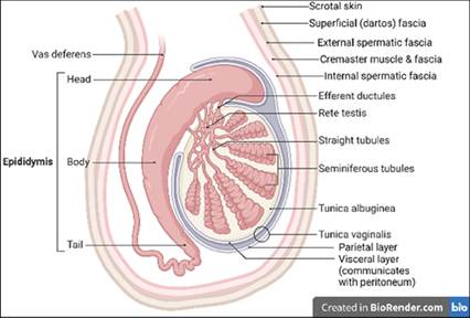
Figure 11.58 Anatomy of the scrotum. The most superficial layer of the scrotal sac is the skin. Deeper facial layers include the superficial (dartos) fascia, the external spermatic fascia, the cremaster muscle and fascia, the internal spermatic fascia, the parietal layer of the tunica vaginalis, and the visceral layer of the tunica vaginalis. Created with BioRender.com.
Neoplasia
In a study of 676 scrotal masses, 96% were neoplastic (Trappler et al., 2014). The vast majority (396 of 676 cases, 58.6%) were round cell tumors; more specifically, 369 of 676 cases (54.6%) were mast cell tumors (Figure 11.59). Melanocytomas were slightly more common than malignant melanomas (7.1% and 4.7%, respectively). Other neoplasms included vascular hamartoma (6.8%), hemangiosarcoma (5.0%), hemangioma (4.6%), and histiocytoma (3.4%). Twenty-nine masses (4.3%) were epithelial in origin; of these, squamous cell carcinoma was the most common (the exact number was not indicated).
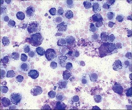
Figure 11.59 Mast cell tumor. FNA from a scrotal mass on a 9-year-old, neutered male American Bulldog. Cells are individualized and round with abundant, lightly basophilic cytoplasm and a large, round, centrally located nucleus. The nuclei have smooth to finely stippled chromatin and an indistinct nucleolus. Cells contain variable numbers of pinpoint metachromatic cytoplasmic granules. Moderate anisocytosis and anisokaryosis are noted. There are occasional mitotic figures. Low numbers of neutrophils and eosinophils, and moderate numbers of erythrocytes are present in the background. Free metachromatic granules are scattered throughout the sample (Wright–Giemsa, 500? magnification).