Squamous cell carcinoma
SCCs tend to exfoliate well. Cells are primarily individualized and round to polygonal with abundant lightly basophilic to aqua cytoplasm. Nuclei are variably sized and round to irregular with coarse to clumped chromatin.
Many classic characteristics of malignancy can be identified. Additionally, aberrant nuclear to cytoplasmic maturation is commonly observed, especially a large nucleus with immature chromatin within a keratinized cell. Perinuclear vacuoles indicative of aberrant keratinization also are common. Tadpole-shaped cells may be seen; these cells have a tail of lightly basophilic cytoplasm and an eccentrically located, rounded nucleus. Cells that contain a large cytoplasmic vacuole that flattens and displaces the nucleus to the side, giving them a signet ring appearance, may also be found (Figures 11.54–11.57). 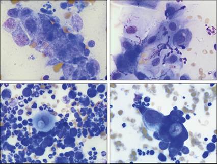
Figures 11.54–11.57 Squamous cell carcinoma. Impression smears from a mass on the penis of an 11-year-old, intact male Dachshund. Figure 11.54: Cells in this image are arranged in a cluster. Cells are large when compared with the neutrophil at the top of the image. They have scant, basophilic cytoplasm and a large, rounded nucleus with finely stippled chromatin and multiple nucleoli. Moderate anisocytosis and anisokaryosis are seen. This morphology supports a diagnosis of carcinoma (Wright–Giemsa, 1,500? magnification). Figure 11.55: Cells in this area of the slide are more individualized and some are keratinized. Cells are large and round to angular with abundant, lightly basophilic cytoplasm and a round nucleus with stippled chromatin. A prominent nucleolus can be seen in low numbers of cells. A few neutrophils also are present (Wright–Giemsa, 1,000? magnification). Figure 11.56: Large numbers of neutrophils and fewer macrophages are seen in some areas of the slide. A single, anaplastic, squamous epithelial cell with abundant light-blue–green cytoplasm, an aberrant cytoplasmic vacuole, and a distorted, rounded nucleus is present at the center of the image (Wright–Giemsa, 1,000? magnification). Figure 11.57: A large cluster of highly variably sized epithelial cells is shown. Cells have scant to abundant, lightly to deeply basophilic, rounded cytoplasm. The nucleus is round with open chromatin. A prominent, crescent-shaped nucleolus is observed in one of the larger cells. Many of the smaller cells have a high nuclear to cytoplasmic ratio. Low numbers of neutrophils are closely associated with the neoplastic cells (Wright–Giemsa, 1,000? magnification).