Cases
CASE 1
Signalment/history
A 4-year-old, neutered male Labrador Retriever was hit by a car 3 days previously and presented for abdominal pain and lethargy.
Physical examination
On physical examination, the patient was febrile and tachycardic with severe abdominal pain.
Abdominal radiographs revealed abundant amounts of peritoneal fluid.Diagnostic tests
A sample was taken and protein and cell concentrations were determined: protein, 3.8 g/dl (38 g/l); nucleated cell count, 6.5 ? 103 cells/μl; red blood cell count, 45 ? 103 cells/μl.
The fluid was characterized as an exudate. Cytologically, a mixed inflammatory population was observed, predominated by neutrophils with lesser numbers of vacuolated macrophages containing a greenish-black pigment suggestive of bile (Figures 15.97, 15.98). The total bilirubin was measured in the effusion and compared with serum: serum total bilirubin, 1.5 mg/dl (25.7 μmol/l); fluid total bilirubin, 6.5 mg/dl (111.2 μmol /l).
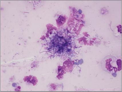
Figure 15.90 Sulfur granule from a patient infected with Nocardia spp. or Actinomyces spp. (Hemafix™, 1,000? magnification).
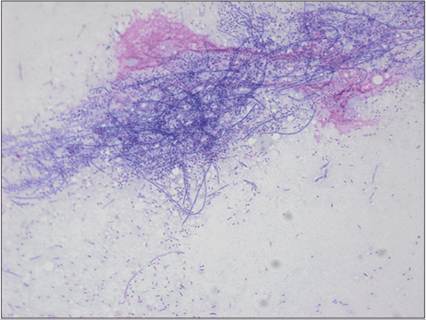
Figure 15.91 A squash preparation of pleural fluid reveals a mold of filamentous septated bacteria. Culture revealed Actinomyces spp. (Hemafix™, 1,000? magnification).
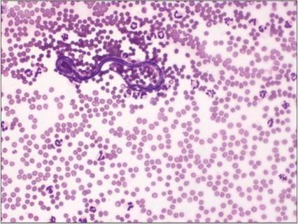
Figure 15.92 Hemorrhagic abdominal fluid from a dog. Rarely, parasites may be observed in samples containing blood. A microfilarial parasite (Dirofilaria repens) is shown in this image.
This patient had a history of blunt abdominal trauma, which caused the hemorrhagic effusion. The organism was an incidental finding in this case (Hemafix™, 400? magnification).
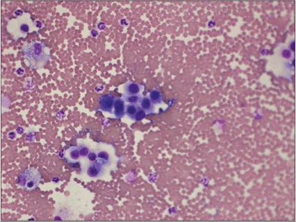
Figure 15.93 Pericardial fluid from a Border Collie with ectopic thyroid carcinoma. Clusters of round, slightly polygonal cells with faint tight junctions, coarse chromatin, and irregular nucleoli (Hemafix™, 400? magnification).
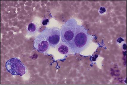
Figure 15.94 Pericardial fluid from a Border Collie with ectopic thyroid carcinoma. The neoplastic cells are large with large prominent nucleoli (Hemafix™, 1,000? magnification).
Figure 15.95 Pleural fluid from a cat with a history of uveal malignant melanoma. Pleomorphic mesenchymal cells exhibiting multiple criteria of malignancy and fine black pigment granules (Hemafix™, 400? magnification).
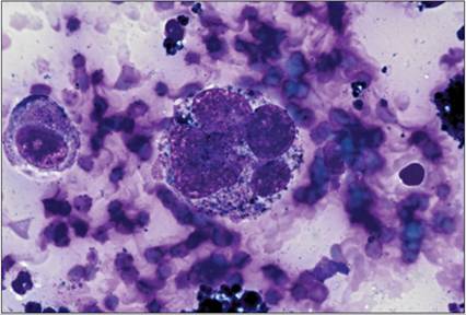
Figure 15.96 Pleural fluid from a cat with a history of uveal malignant melanoma. Multinucleated giant cell containing blue–black melanin granules (Hemafix™, 1,000? magnification).
Figure 15.97 Abdominal fluid from the patient presented in case 1. The sample is cellular and consists of neutrophils with fewer macrophages containing pigmented material within the cytoplasm (Hemafix™, 400? magnification).
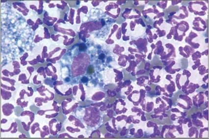
Figure 15.98 Within the cytoplasm of the macrophage, pigmented material consistent with bile is visualized (Hemafix™, 1,000? magnification).
Diagnosis
Bile peritonitis.
Management
An emergency exploratory laparotomy was performed, and a ruptured gallbladder was identified and removed.
CASE 2
Signalment/history
A female spayed Bichon Frisè, 11 years old, was on a hypoallergenic diet because of chronic diarrhea varying in severity suspected of inflammatory bowel disease. She was referred because of labored breathing for 2 days and coughing. The dog has been eating well most of the time; now she has no appetite.
Physical examination
On physical exam, body condition score 2/5; muffled heard sounds, abdominal distention with undulation. Diagnostic imaging: liquidothorax and mild ascites. Gastric and intestinal biopsies were taken by endoscopy.
Diagnostic tests
Bloodwork revealed mild thrombocytosis, neutrophilia, increased C-reactive protein (102 mg/L; RI: mirror-like cytoplasmic protrusions. Note the mitosis top right (Hemafix™, 400? magnification).
Figure 15.111 Pleiomorphic lymphoid round cells and cytoplasmic protrusions (Hemafix™, 400? magnification).
Figure 15.112 Pleiomorphic lymphoid round cells and cytoplasmic protrusions and clod-like nuclear chromatin (Hemafix™, 1,000? magnification).