Chemical Basis for Life[*]
Joanna M. Bassert
OUTLINE
INTRODUCTION, 12
MATTER, 13
States of Matter, 13
Composition of Matter: Elements and Atoms, 13
Elements, 13
Atoms, 15
Molecules and Compounds, 19
Mixtures, 19
Distinguishing Compounds from Mixtures, 21
CHEMICAL BONDS, 21
Covalent Bonds, 21
Ionic Bonds, 21
Hydrogen Bonds, 23
Chemical Reactions, 24
CHEMICAL COMPONENTS OF LIVING ORGANISMS, 25
INORGANIC COMPOUNDS, 26 Water, 26
Salts, 28
Acids and Bases, 28
ORGANIC COMPOUNDS, 31
Carbohydrates, 31
Lipids, 34
Proteins, 35
Nucleic Acids, 40
LEARNING OBJECTIVES
When you have completed this chapter, you will be able to:
1.
List the characteristics of each of the subatomic particles.2. Differentiate between a molecule and a compound.
3. List and describe the types of chemical bonds that may form between elements.
4. Give the general equations for synthesis, decomposition, and exchange reactions.
5. Differentiate between organic and inorganic compounds.
6. Differentiate between hydrophobic and hydrophilic molecules.
7. List the unique properties of the water molecule.
8. Differentiate between acids and bases.
9. Describe the actions of a buffer system.
10. List the components of carbohydrates, lipids, proteins, and nucleic acids.
11. List the functions of body proteins.
12. Describe the actions of enzymes.
VOCABULARY FUNDAMENTALS
Acid ah-sihd
Activation energy ahck-tuh-va-shuhn ehn-ar-je
Adenosine diphosphate ah-dehn-o-sen dι-fohs-fat Adenosine triphosphate ah-dehn-o-sen trι-fohs-fat Amino acid ah-me-no ah-sihd
Anion ahn-ι-uhn
Atom aht-uhm
Atomic number ah-tohm-ihck nuhm-bar
Atomic weight ah-tohm-ihck wat
Base bas
Carbohydrate kahr-bo-hι-drat
Catalyst kaht-ah-lihst
Cation kaht-ι-ohn
Cellular respiration sehl-u-lar res-puh-ra-shuhn Chemical element kehm-ih-kuhl ehl-uh-mehnt Chemical equation kehm-ih-kuhl e-kwey-shuhn Chemical reaction kehm-ih-kuhl re-ahck-shuhn Chemical symbol kehm-ih-kuhl sihm-buhl Chromosome kro-mo-som
Colloid kohl-oyd (or emulsion e-muhl-shuhn) is a heterogeneous heht-ar-rohj-uh-nuhs mixture mihcks-char that contains a much larger sized
solute sohl-yoot than those found in a
solution suh-loo-shuhn
Compound kohm-pohwnd
Covalent bond ko-va-lehnt bohnd
Decomposition reaction de-kohmp-o-zihsh-uhn re-ahck-shuhn
Dehydration synthesis de-hι-dra-shuhn sihn-thuh-sihs
Deoxyribonucleic acid (DNA) de-ohck-se-rι-bo-noo-kla- ihck ah-sihd
Disaccharide dι-sahck-uh-rιd
Eicosanoid ι-ko-seh-noyd
Electron e-lehck-trohn
Electron shell e-lehck-trohn shehl
Electrostatic attraction e-lehck-tro-staht-ihck ah-trahck-shuhn
Element ehl-eh-mehnt
Enzyme ehn-zιm
Exchange reaction ehcks-chanj re-ahck-shuhn
Fatty acid faht-e ah-sihd
Functional group fuhngk-shuh-nuhl groop
Functional protein fuhngk-shuh-nuhl pro-ten
Glycerol glihs-or-ahl
Glycoprotein glι-ko-pro-ten
Hydrolysis hι-drohl-uh-sihs
Hydrophilic hι-dro-fihl-ihck
Hydrophobic hι-dro-fo-bihck
Inorganic compound ihn-ohr-gahn-ihck kohm-pohwnd
Ion ι-ohn
Ionic bond ι-ohn-ihck bohnd
Isotope ι-so-top
Lipid lihp-ihd
Lipoprotein lι-po-pro-ten
Macromolecule mah-kro-mohl-uhl-kyool
Matter maht-or
Mixture mihcks-chor
Molecule mohl-uhl-kyool
Monosaccharide mohn-o-sahck-ah-rιd
Neutral fat noo-truhl faht
Neutralize noo-truhl-ιz
Neutron noo-trohn
Nucleotide noo-kle-o-tιd
Organic compound ohr-gahn-ihck kohm-pohwnd
Peptide bond pehp-tιd bohnd
Periodic Table of the Elements peer-e-ohd-ihck ta-buhl of the ehl-eh-mehntz
Phospholipid fohs-fo-lihp-ihd
Polypeptide pohl-e-pehp-tιd
Primary structure prι-meor-e struhckt-shor
Product prohd-uhckt
Protein pro-ten
Proton pro-tohn
Radioactive isotope rad-e-o-ahck-tihv
ι-so-top
Reactant re-ahck-tuhnt
Ribonucleic acid (RNA) rι-bo-noo-kla-ihck
ah-sihd
Salt sahlt
Saturated fatty acid sahch-or-a-tihd faht-e ahs-ihd
Solute sohl-yoot
Solution suh-loo-shuhn
Solvent sohl-vuhnt
Steroid stear-oyd
Structural protein struhck-shor-uhl pro-ten
Substrate suhb-strat
Suspension suh-spehn-shuhn
Synthesis reaction sihn-thuh-sihs re-ahck-shuhn
Triglyceride trι-glihs-or-rιd
Unsaturated fatty acid uhn-sahch-or-a-tihd
faht-e ah-sihd
INTRODUCTION
When our universe was less than 1 second old, astronomers tell us, waves of hydrogen and energy streamed across an ever-expanding space.
Gravity swirled these small atoms closer and closer until they were crushed against one another, igniting the nuclear reactions that formed the stars. The products of the nuclear reactions were new elements that were blown into the cosmos when stars exploded into supernova. Oxygen, nitrogen, iron, silicone, and other elements were forged in these nuclear furnaces. Carbon was blown off of slowly aging, mediumsized stars and condensed into minute dust particles. Four and a half billion years ago the debris from these stellar explosions coalesced by the force of gravity to form celestial bodies that circled our sun. Earth was created literally from stardust (Figure 2-1).The atmosphere of the primordial earth contained methane gas (CH4), water (H2O), and ammonia (NH4) but little free oxygen. These molecules contain the
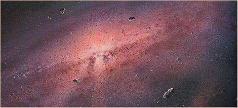
FIGURE 2-1 Illustration of the early cosmos. A painting of early events in the universe (NASA/JPL). (From Weissman P, McFadden L, Johnson T: Encyclopedia of the solar system, Burlington, Mass, 1999, Academic Press.)
elements — hydrogen, oxygen, carbon, and nitrogen—that make up 96% of living organisms. Scientists believe that the combined activity of lightning, ultraviolet (UV) light, meteorite strikes, and thermo-reactions in the earth's crust and core provided the energy needed to convert CH4, H2O, and NH4 into life-generating organic molecules such as amino acids and nucleic acids. The most successful molecules were self-replicating. From these self-replicating molecules, scientists believe early cells evolved. These early cells were tiny, bacteria-like units without a nucleus, and they had the new ability to use energy from the environment to make their own chemical energy.
Archaebacteria are ancient bacteria that survived the harsh, oxygen-free environment of young earth. Some forms still exist today in the extreme environments of hot springs, salt flats, and the intestines of mammals.
Early bacteria had various ways of creating the chemical energy needed to maintain themselves and to reproduce. Some developed enzymes that created oxygen gas as a waste product and, later, cells evolved that used oxygen to produce their own molecular energy. As oxygen concentrations in the earth's atmosphere climbed, expansion of the number and size of living organisms became possible (Figure 2-2).Life on earth comes in a multitude of forms, but the biochemistry that defines living things is remarkably consistent. All living entities are formed from inorganic chemicals, such as water and salts, as well as from organic chemicals, such as proteins, lipids, carbohydrates, and nucleic acids. The same physical forces that caused the formation of the stars, planets, and elements also governed biochemical reactions and interactions in living organisms.
The body of an animal is composed of thousands of chemicals, interacting with one another at rapid speed. Their dynamic collisions with and separations from one
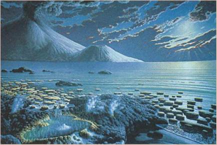
FIGURE 2-2 The Precambrian sea. The earliest evidence of life is shown by calcified algal structures called stromatolites that existed during the Precambrian period. Scientists believe that the energy from asteroid strikes, lightning strikes, and the reactions in the earth's melted crust and core supplied the energy for common molecules, such as ammonia and methane, to be converted into organic molecules such as proteins and nucleic acids. (Courtesy the National Museum of Natural History, Smithsonian Institution.)
another underlie the physiological processes of life: respiration, digestion, reproduction, movement... they are all the result of chemical interaction. Each organic and inorganic molecule found in living systems is composed of atoms, the elemental units of matter. It is fitting, therefore, to begin our discussion of anatomy and physiology with an introduction to biochemistry, the chemistry of life.
MATTER
Matter is defined as anything that occupies space and has mass (Figure 2-3).
We can often identify matter with our senses by feeling, seeing, tasting, and smelling. Though matter has mass and takes up space, keep in mind that mass is not the same as weight. The mass of an animal is based on how much matter it contains, whereas an animal's weight is determined by the pull of gravity on the matter. On earth, for example, a unit of matter would weigh more than the same unit of matter on the moon, because the acceleration of gravity on the moon is far less than that on earth. However, the mass is the same on both the earth and the moon.STATES OF MATTER
Matter can exist in one of three states, as a gas, liquid, or solid. The bodies of animals contain examples of each state. The air that is inhaled and the carbon dioxide that is exhaled are examples of gases in the living system. Blood, which is primarily composed of water, is a vital liquid that helps transport critical nutrients to hungry tissues. Finally, the musculoskeletal system composed of bones, tendons, ligaments and muscles are examples of solid features that give the body shape and strength.
COMPOSITION OF MATTER:
ELEMENTS AND ATOMS
ELEMENTS
All matter is made of one or more elements. Each element is a single pure substance consisting of only one type of atom. All of the 118 elements known today are listed in the Periodic Table of Elements where they are divided into three general categories: metals, metalloids, and nonmetals (Figure 2-4). Only 92 of the elements occur in nature; the rest are made artificially or are theoretical and not known to exist.
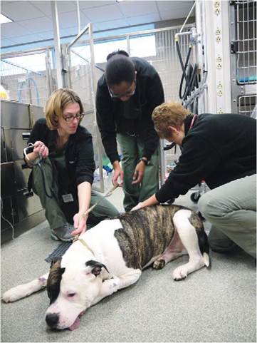
FIGURE 2-3 Matter. Everything you see in this picture is made of matter. Matter is anything that takes up space and has mass. The page it is printed on is also matter. (Courtesy Dr. Joanna Bassert.)
Each known element has its own unique properties, but it can be joined in various combinations with other elements to form all of the matter that exists on earth.
Some common examples of elements are aluminum, gold and carbon as pictured in Figure 2-5. Others include oxygen, chlorine, and helium. At room temperature, gold is a solid metal, whereas oxygen and chlorine are gases. Pure carbon can exist as coal or, with enough time and pressure, can be compressed into diamonds.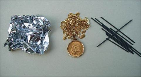
FIGURE 2-5 Exampl es of elements. Aluminum foil is 98.5% aluminum.
Twenty-four carat gold is 99.9% gold. Graphite is 100% carbon.
Periodic Table
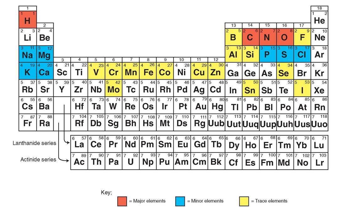
FIGURE 2-4 Periodic Table of the Elements. The Periodic Table of the Elements gives us important information about each element: the chemical symbol, atomic number, and atomic weight. The table groups elements with similar properties. The metallic elements are on the left and the inert gases are in the right-hand column. The elements shaded in red are the major elements that make up 96% of the matter in the animal body. The elements shaded in blue are the minor elements, and those shaded in yellow are trace elements.
Surprisingly, living organisms are made up of only a few of the 118 known elements. Only four elements, nitrogen, oxygen, hydrogen, and carbon, make up 96% of the matter found in all living organisms. Several other elements are found in relatively small quantities. Table 2-1 shows the most common elements found in living organisms and their function in the body. These are divided into major and minor categories.
Each element is referenced using a chemical symbol, which is derived from its name in English, Latin, or Greek. The chemical symbol for oxygen, for example, is O from the English word “oxygen,” whereas the chemical symbol
for gold is Au from the Latin word aurum, meaning gold (Figure 2-4).
ATOMS
An atom is the smallest unit of an element that retains the unique properties of the element (Figure 2-6).
Atoms themselves are made of smaller, subatomic particles called protons, neutrons, and electrons, but these particles do not retain the physical and chemical properties of the element when they are isolated. The protons and neutrons are the heaviest particles—each has an atomic mass of one—and these are grouped together in the center of the atom and collectivelyElements in the Animal Body
The Percentage of Each Element Found in an Animal's Body is Listed. Note That the First Few Elements Make Up the Vast Majority of Matter in the Animal Body
TABLE 2-1
| ELEMENT | CHEMICAL SYMBOL | ATOMIC NUMBER | BODY MASS (%) | FUNCTION IN THE ANIMAL BODY |
| Major Elements | ||||
| Oxygen | O | 8 | 65.0 | Necessary for cellular respiration; component of water |
| Carbon | C | 6 | 18.5 | Primary component of organic molecules |
| Hydrogen | H | 1 | 9.5 | Component of water and organic molecules; necessary for energy transfer and respiration; ion influences pH of fluids |
| Nitrogen | N | 7 | 3.3 | Component of all proteins and nucleic acids |
| Calcium | Ca | 20 | 1.5 | Component of bones and teeth; required for muscle contraction, nerve impulse transmission, and blood clotting |
| Phosphorus | P | 15 | 1.0 | Principal component in backbone of nucleic acids; important in energy transfer (part of ATP); component of bones |
| Potassium | K | 19 | 0.4 | Principal positive ion within cells; important in nerve function |
| Sulfur | S | 16 | 0.3 | Component of most proteins |
| Sodium | Na | 11 | 0.2 | Important positive ion in extracellular fluid; important in nerve function |
| Chlorine | Cl | 17 | 0.2 | Ion is most abundant negative ion in extracellular fluids |
| Magnesium | Mg | 12 | 0.1 | Component of many energy-transferring enzymes |
| Trace Elements | ||||
| Silicone | Si | 14 | 0.1 | Component of some enzymes |
| Aluminum | Al | 13 | 0.1 | Component of some enzymes |
| Iron | Fe | 26 | 0.1 | Critical component of hemoglobin |
| Manganese | Mn | 25 | 0.1 | Needed for fatty acid synthesis |
| Fluorine | F | 9 | 0.1 | Component of bones and teeth |
| Vanadium | V | 23 | 0.1 | Component of some enzymes |
| Chromium | Cr | 24 | 0.1 | Needed for proper glucose metabolism |
| Copper | Cu | 29 | 0.1 | Needed for hemoglobin and myelin |
| Boron | B | 5 | 0.1 | Component of some enzymes |
| Cobalt | Co | 27 | 0.1 | Needed for maturation of red blood cells |
| Zinc | Zn | 30 | 0.1 | Important component of many enzymes and proteins |
| Selenium | Se | 34 | 0.1 | Antioxidant |
| Molybdenum | Mo | 42 | 0.1 | Key component of many enzymes |
| Tin | Sn | 50 | 0.1 | Component of some enzymes |
| Iodine | I | 53 | 0.1 | Component of thyroid hormones |
From Patton KT, Thibodeau GA: Anatomy and physiology, ed 8, St Louis, 2013, Mosby, p 35.
∕ j CLINICAL APPLICATION
Iron Deficiency Anemia
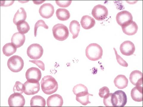
There are trace amounts of some elements in the body that are essential for life. Iron is an example of an essential element. As a percentage of the mass of the body, iron exists in extremely small amounts. Healthy animals have only 9 to 22 mg of iron in their bodies, most of which is found in the globular protein, hemoglobin, in red blood cells. Iron is used to bind oxygen and carry it to tissues where it is needed in the mitochondria to generate adenosine triphosphate (ATP). Without iron, the level of oxygen that can be carried by the blood is reduced, leading to fatigue and exercise intolerance. Chronic blood loss reduces the level of iron stored in the body. Without adequate levels of iron, the body is not able to make hemoglobin and adequate numbers of red blood cells. In this way, ongoing blood loss results in a condition called iron deficiency anemia. A puppy or kitten with a severe flea infestation, for example, can lose 100 ml of blood a day. When the stores of iron in the body are depleted, hemoglobin can no longer be manufactured and the red blood cell count decreases. Clinical signs include pale mucous membranes, fatigue, bounding pulses, and galloping heart rhythm. Microscopic examination of the blood shows small (microcytic) pale (hypochromic) red blood cells with large central areas. The oxygen carrying capacity of blood affected by iron deficiency is greatly reduced and the animal becomes weak. If the animal is stressed and the demand for oxygen increases, such as during a physical examination at the vet’s office, the puppy or kitten can die suddenly from heart failure. Iron deficiency anemia is treated by stabilizing the patient with blood transfusions, giving oral and injectable iron supplements, and eliminating the cause of the blood loss.
From Harvey JW: Veterinary hematology: a diagnostic guide and color atlas, St Louis, 2012, Elsevier Saunders.
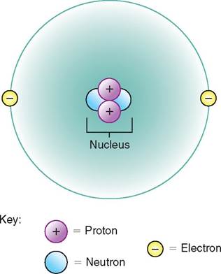
FIGURE 2-6 Diagram of a helium atom (planetary model). The neutrons and protons group together in the center of the atom, which is called the nucleus. Electrons move around the outside of the nucleus in an orbit. The charges of the particles are shown. Note that there are equal numbers of protons and electrons, giving the atom no net electrical charge.
are called the atomic nucleus. The neutrons and protons together determine the atomic weight of the atom. Electrons are tiny “wavicles” that possess the properties of both waves and particles. They can collide with other electrons and can be diffracted like light. Electrons exist in a state of constant motion moving continuously around the nucleus and
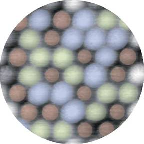
FIGURE 2-7 Electron Clouds. Using an atomic force microscope (AFM), the outer surfaces of electron clouds are visible in atoms clustered together along the flat surface of a crystal. The different colors represent different types of atoms. (From Sugimoto Y, Pou P, Abe M, et al: Chemical identification of individual surface atoms by atomic force microscopy, Nature 466:64-67, 2007.)
generate regions where they are statistically most likely to be found called electron clouds (Figure 2-7). Electrons are so tiny that their mass does not contribute to the atomic weight of the atom as a whole. Protons have a positive electrical charge, electrons have a negative electrical charge, and neutrons have no electrical charge and are therefore neutral. The net electrical charge of many atoms is neutral, because the atom contains equal numbers of protons and electrons, so the positive and negative charges of these particles cancel each other out.
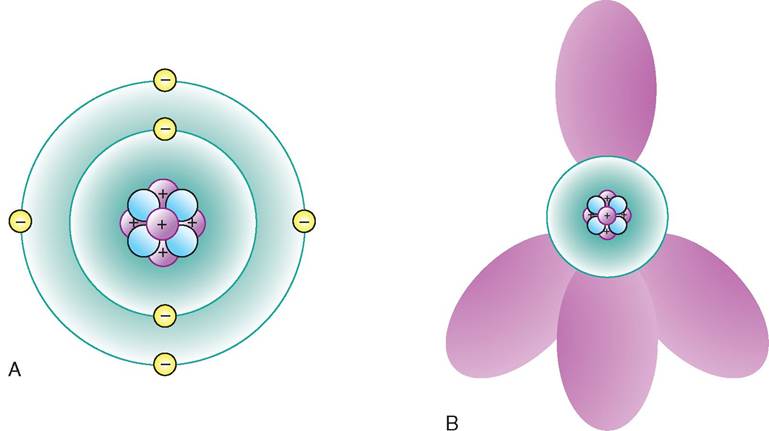
FIGURE 2-8 Atomic models. A, The planetary model of an atom of carbon. Carbon has six protons, six electrons, and six neutrons. (Note: Not all the nuclear particles are shown.) B, An orbital model of carbon. The neutrons and protons are grouped together in the nucleus. The shaded shapes show the most likely positions of the various electrons that surround the nucleus.
There are two common models used to represent atoms graphically. The planetary model shows the protons and neutrons of the nucleus encircled by electrons, which orbit like planets around a sun. This is not physically accurate, but it makes the atom easy to understand and allows us to explain interactions between two atoms in a clearer fashion. A more accurate representation is the orbital model, which shows a three-dimensional view of the most likely location of the electrons at any given time. Figure 2-8 shows the difference between a planetary model and an orbital model of a carbon atom. Keep this model in mind while studying the interaction of atoms and molecules. Electrons exist in a cloud around the nucleus, and can move closer to one side of the nucleus than another. Each atom of an element has the same number of protons as other atoms of that element. For example, every oxygen atom has eight protons, and every carbon atom has six protons. If the number of protons is changed—which can only be done with extraordinary means, like nuclear reactions—then the element is changed to another element. In each atom, the number of electrons is the same as the number of protons. The atomic number of the element is equal to the number of protons found in the nucleus. Sometimes an atom can lose or gain an electron, giving it a positive or negative charge: This charged atom is called an ion. Each element has a naturally occurring, specific number of neutrons. Figure 2-8 shows examples of common elements that make up the animal body.
Sometimes atoms exist that contain a different number of neutrons. These atoms are called isotopes of the element. For example, carbon normally has six protons, six electrons, and six neutrons, so the atomic weight of carbon is normally 12. There are some isotopes of carbon, however, that have eight neutrons, giving them the atomic weight of 14. This
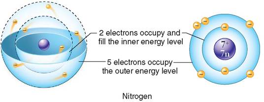
FIGURE 2-9 The principal quantum numbers or energy levels of the electron shells of a nitrogen atom are depicted in three dimensions (left) and using a planetary model (right). (From Patton KT, Thibodeau GA: Anatomy and physiology, ed 8, St Louis, 201 3, Mosby, p 37, Figure 2-5.)
form of carbon is a radioactive isotope called carbon-14. A radioactive isotope spontaneously emits particles of energy at a constant rate and thereby changes into a stable, nonradioactive element. The rate at which this happens is called the rate of decay. Carbon-14's rate of decay can be measured in rock and is used to date fossils.
The area around the nucleus where the electrons have their most likely position is called the electron shell. An electron's energy level determines which electron shell it will inhabit. An atom has one or more electron shells surrounding the nucleus, depending on the number of electrons and their energy. Lower energy electrons exist in the first electron shell, which is found closest to the nucleus. Higher energy electrons are in the second electron shell (Figure 2-9).
We will use the planetary model to describe the electron shell (Figure 2-10). The first electron shell can only hold two electrons. Hydrogen has only one electron, so it has only one electron shell. Helium has two electrons, so its first and only
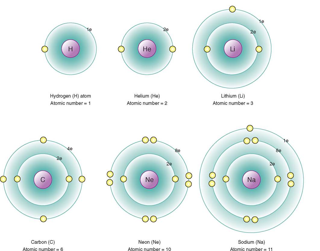
FIGURE 2-10 Diagram of atoms showing the electron shells. Note that the outer shells of hydrogen, lithium, carbon, and sodium are incomplete, but the outer shells of helium and neon are complete. This renders the atoms of helium and neon less chemically active; indeed, they are referred to as inert gases.
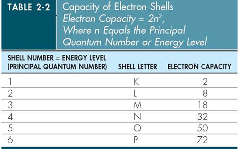
electron shell is full. The next element in the Periodic Table is lithium, which has three electrons; so the first electron shell is full, and the second electron shell contains one electron. The second electron shell can hold up to eight electrons. Carbon has six electrons, so its first shell is full, and its second shell contains four electrons. Neon has 10 electrons, so its first and second shells are completely filled. It is important to understand whether the electron shell of a particular atom is full or incomplete. Atoms are most stable when their electron shells are full. Helium and neon have full electron shells and are therefore chemically inactive or inert. Atoms like hydrogen, carbon, and oxygen are more chemically active, because their electron shells are incomplete. These atoms are constantly trying to find electrons to complete their outer shell (Table 2-2). The forces that drive the activities of electrons account for the formation of bonds between atoms.
MOLECULES AND COMPOUNDS
A molecule forms when atoms are joined together by chemical bonds. If two or more atoms of the same element are joined together, we call the result a molecule of the element. For example, oxygen does not usually exist as a single atom. Oxygen gas exists as a molecule of two oxygen atoms joined together, and this is expressed by the symbol O2 (Figure 2-11). The subscript 2 denotes the number of atoms in the molecule.
Atoms of different elements may also join together to form a molecule. For example, carbon dioxide is formed from one atom of carbon and two atoms of oxygen. The chemical symbol for carbon dioxide is CO2 (Figure 2-12). A molecule of table salt is called sodium chloride and has the chemical symbol NaCl. A molecule of this compound consists of one atom of sodium bonded to one atom of chlorine (Figure 2-13). A molecule is the smallest unit of a compound that retains the properties of that compound. This is important because often the properties of the compound are much different than the properties of the elements from which it is made. Table salt is very different from sodium (a metal) and chlorine (a poisonous gas).
MIXTURES
Most matter is combined into mixtures of two or more substances. There are three types of mixture: solutions, colloids, and suspensions (Figure 2-14).
Solutions are homogeneous mixtures of various substances. The components can be gases, liquids, and/or solids. A component that is present in the greatest amount is called the solvent, while the substances present in smaller amounts
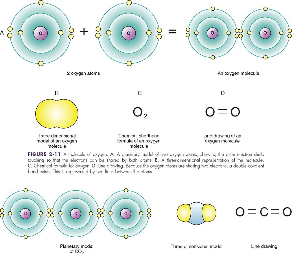
FIGURE 2-12 A molecule of carbon dioxide. Methods of representing a molecule of carbon dioxide (CO2) are shown. The carbon atom is sharing two electrons with each of the oxygen atoms to form double covalent bonds.
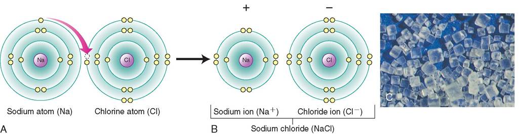
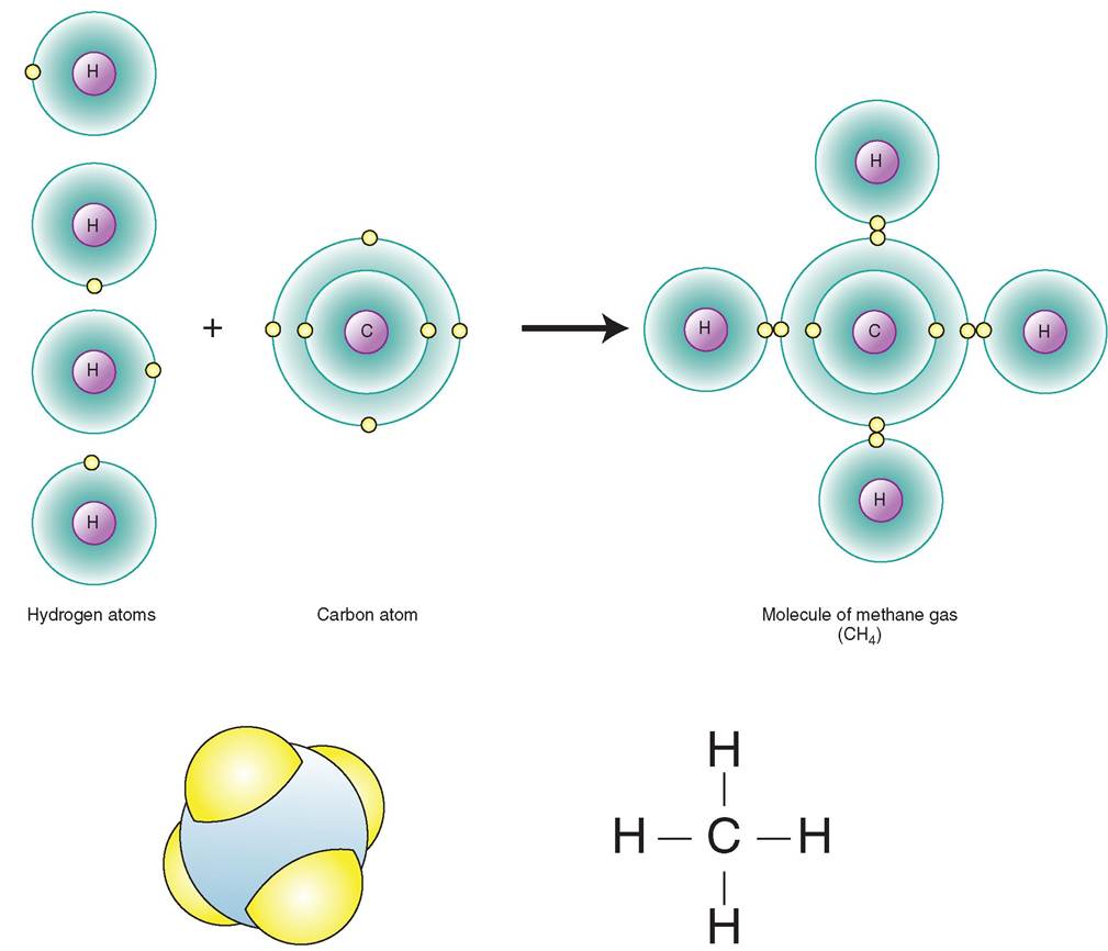
FIGURE 2-13 Sodium chloride (NaCl) is formed from ionic bonding. A, Within ⅛e aqueous environment of the body, ionic: bonds are frequently formed as electrons are transferred from sodium ions to chloride ions. This gives both atoms an electrical charge. B, Table salt takes on cube-shaped crystals. C, A photomicrA single covalent bond is formed when one electron is shared. When two electrons are shared the bond is called a double covalent bond. A triple covalent bond is formed when three electrons are shared (Figure 2-15).
The shared electrons in a covalently bonded molecule spend part of their time in the electron shell of each of the atoms. Sometimes the electrons spend more time in one atom than in the other. For example, the water molecule H2O is made of two hydrogen atoms and one oxygen atom. The shared electrons spend more time near the oxygen atom because oxygen is a good electron acceptor and hydrogen is a good electron donor (Figure 2-16). Because of this distribution of electrons and the three-dimensional arrangement of the molecule, there will be a slight positive charge on the hydrogen side of the molecule and a slight negative charge on the oxygen side. The position of the covalent bonds in water arranges the hydrogen atoms toward the same side of the oxygen molecule. This makes the molecule a polar molecule, meaning it has oppositely charged ends. This special electromagnetic property of water will be important in the interactions of many molecules in the body. We will explore this further when we discuss the properties of water.
IONIC BONDS
An ionic bond is formed when electrons are transferred from one atom to another. Ionic bonds are most often formed between two types of atom: those with fewer than two electrons in their outer shell and those with nearly full outer shells. An atom with one electron in its outer shell will be inclined to give up that electron so its “new” outer shell will be stable. Similarly, an atom that needs only one electron readily accepts electrons that will make its outer shell full and stable. For example, the sodium atom (Na) has 11 electrons: two in the first shell, eight in the second shell, and one in the
Three dimensional model Line drawing
FIGURE 2-15 Formation of single covalent bonds. Planetary model, three-dimensional model, and line drawing of a methane molecule (CH4). Since each hydrogen atom is sharing only one electron with the carbon atom, a single covalent bond is formed, represented by a single line.
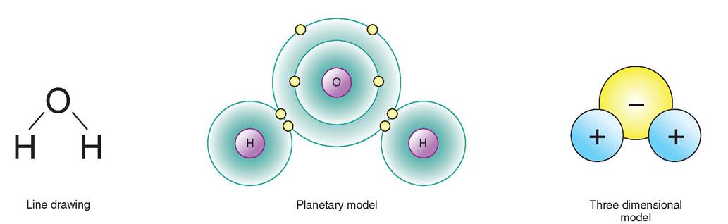
FIGURE 2-16 The polar water molecule. Bonded electrons spend more time near the oxygen atom, giving that aspect of the molecule a slightly negative charge. The hydrogen side of the molecule carries a slightly positive charge.
third shell. It would be more stable without the 11th electron. Suppose a handy atom like chlorine is nearby, with 17 electrons: two in the first shell, eight in the second shell, and seven in the third shell. It badly needs one more electron to make its third shell stable (see Figure 2-13). As the atoms approach each other, sodium's outer electron is strongly attracted to the third shell of the chlorine atom. This does a few things: When the sodium atom loses its electron, it develops a positive charge, because it has more protons than electrons. When the chlorine atom gains the extra electron, it gains an overall negative charge, because now it has more electrons than protons. Thus, these two atoms are drawn to each other by their respective electrical charges, which is an electrostatic attraction. This is an ionic bond, and the resulting compound is a molecule of sodium chloride, or table salt (NaCl) (Figure 2-17). Sodium and chloride are ions because they have an electrical charge. Sodium is a cation because its electrical charge is positive. All cations have positive charges. You can remember this if you think of the “T” in “cation” as a “+” sign. For those of you who really like cats, you will not find it difficult to remember that cations are positively charged ions. Chloride has a negative charge and is called an anion. All anions have a negative charge. All ions (cations and anions) are involved in essential functions in the animal body including, for example, contraction of muscle fibers, transmission of nerve impulses, and maintenance of water balance.
HYDROGEN BONDS
A hydrogen bond is more of an electrostatic attraction than a true bond because electrons are neither shared nor transferred. It is far weaker than either a true ionic bond or a covalent bond, and is formed when a hydrogen atom (that is already covalently bonded to an atom) is electrostatically attracted to another hydrogen atom that is covalently bonded to a separate atom on a separate portion of the same molecule or on a separate molecule altogether. When hydrogen is covalently bonded in a molecule, it usually has a slight positive charge. As we have already discussed, hydrogen is a willing electron donor, so the shared electron spends more time away from the hydrogen atom, giving it a relative positive charge. A good example of hydrogen bonding occurs in water (H2O). In this molecule, the hydrogen atoms' electrons are electrostatically attracted toward the oxygen atom. This gives the hydrogen side of the molecule a slight positive charge, so the hydrogen side is subsequently attracted by electrostatic forces to the negatively charged portion (the oxygen side) of other water molecules (Figure 2-18). Hydrogen bonds are formed mostly between molecules— such as between water molecules—and act to stabilize the solution. Hydrogen bonding is key to water's unique properties as a universal solvent and medium for life processes. Hydrogen bonds also can form between parts of the same molecule,lhpwisch he to stabilize the molecule. The shape of large complex molecules, such as proteins and DNA, is maintained by hydrogen bonding within the macromolecule (Figure 2-19).
TEST YOURSELF 2-2
1. What is a molecule?
2. How does an ionic bond differ from a covalent bond?
3. In what circumstance does a hydrogen bond commonly occur?
4. What important function do hydrogen bonds perform in organic and inorganic chemicals?
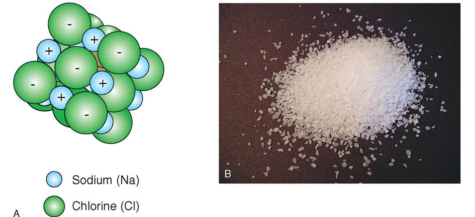
FIGURE 2-17 Ion ic bonding. A, The positive charge of the sodium ion and the negative charge of the chloride ion hold the molecule together by electrostatic attraction, forming ionic bonds. B, The molecules of sodium chloride are also held together by electrostatic attraction, creating the solid form of sodium chloride called table salt.
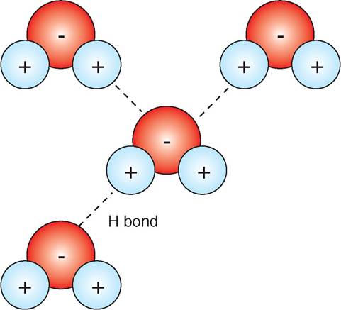
FIGURE 2-18 Hydrogen bonding between water molecules. In this diagram of water molecules, note that the positively charged hydrogen atom of one water molecule is attracted to the negatively charged oxygen atom of another water molecule.
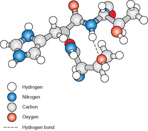
FIGURE 2-19 Hydrogen bonding within protein molecules. In this protein molecule, note the attraction between the hydrogen atom in one part of the molecule and the negatively charged oxygen atom in another portion of the same molecule. Hydrogen bonds hold the protein molecule in its specific shape.
CHEMICAL REACTIONS
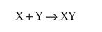
A chemical reaction involves the formation and breaking of chemical bonds. A chemical equation is the way in which the reaction is described in writing; it shows the molecular formula of the reactants (X and Y), the products (Z) and the direction of the reaction (shown by an arrow).
X + Y → Z
Surprisingly, there are only three types of chemical reaction: synthesis, decomposition, and exchange reactions (Figure 2-20).
During a synthesis reaction a new and more complex chemical is made by combining multiple smaller molecules or elements together. Bonds are formed in all synthesis reactions.
For example, the formation of the oxygen molecule (O2) can be written:
Synthesis reactions occur in the body when simple molecules such as amino acids are assembled to form larger peptide chains, which in turn can be assembled to form proteins needed by the body. Synthesis reactions underlie all anabolic (constructive) processes and are particularly evident during growth and the repair of tissues.
In a decomposition reaction a single chemical is broken down into multiple, smaller, chemical units.
An example is the breakdown of water into hydrogen and oxygen gas.
Notice that the number used as a subscript to an element denotes the number of atoms of the element in the molecule (e.g., H2). The number used as a prefix to a molecule denotes the number of molecules of reactant used or of product created (e.g., 2H2O).
Decomposition reactions are the reverse of synthesis reactions and involve the breakdown of chemical bonds. Decomposition reactions are the foundation of catabolic (degradative) reactions. Proteins for example are degraded into smaller peptide chains or even further into individual amino acids. Decomposition reactions are particularly conspicuous during digestion.
In an exchange reaction certain atoms are exchanged between molecules. It is a combination of a synthesis and decomposition reaction. Bonds are both broken and made. In an exchange reaction, new molecules are produced when chemical partners are exchanged.

An example is the reaction of sodium bicarbonate, often given for relief of indigestion, with hydrochloric acid in the stomach: 
Note that the number of atoms of each element is the same on both sides of all chemical equations.
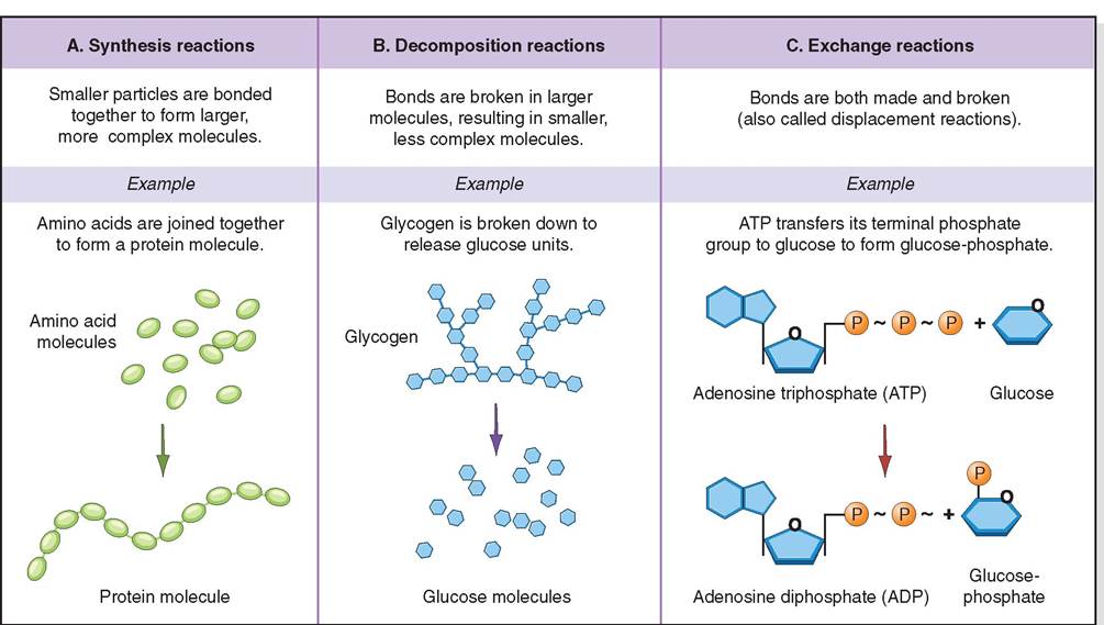
FIGURE 2-20 Three types of chemical reaction: A, synthesis, B, decomposition and C, exchange.
Chemical reactions either require the input of energy or they release energy. In synthesis reactions new bonds are formed, so energy is required. After the bonds are formed, potential energy is stored in the chemical bonds between the atoms. In decomposition reactions energy is released from the breaking of the chemical bonds (i.e., the potential energy stored in the bonds is released). Exchange reactions have no net energy requirement. The energy released from breaking bonds is used to create the new bonds. This concept will be explored in more detail in Chapter 17, Nutrients and Metabolism.
Several factors can influence the rate of reaction. One is the availability of the reactants, referred to as the concentration of reactants. The more reactants that are available, the more likely they will come in contact and be able to react with one another. The temperature of the environment influences the rate of reaction. When the temperature increases, the speed of molecular movement increases, and the chance of molecules meeting improves. Temperature also increases the velocity at which reactants meet, and the velocity provides the energy for the reaction. Activation energy is the energy required for the reaction to happen. Some reactions have a higher activation energy and require the input of more energy for the reaction to occur; these reactions will occur at a slower pace. Certain reactions require the presence of a catalyst. In living organisms, catalysts are usually special proteins that hold the reactants together so they may interact. The catalyst protein is not destroyed or used up by the reaction, and the reaction speed is increased when there are more catalyst proteins present. These special catalyst proteins are called enzymes.
TEST YOURSELF 2-3
1. What is a chemical reaction?
2. What are the three types of chemical reaction?
3. The digestion of food uses which type of chemical reaction?
4. What factors influence the rate of chemical reactions?
CHEMICAL COMPONENTS OF LIVING ORGANISMS
With the myriad of chemical elements available on earth and the millions of combinations of those elements, it is amazing that all living organisms are composed of only a few elements. The compounds that make up living organisms such as a cow, for example, are divided into two categories: organic and inorganic (Figure 2-21). Organic compounds tend to be large, complex molecules that contain carbon-carbon (C-C) covalent bonds or carbonhydrogen (C-H) covalent bonds. Examples of organic molecules include proteins, carbohydrates, triglycerides, and nucleic acids. Inorganic molecules, such as water, salts, acids, and bases, rarely contain carbon and do not contain C-C or C-H bonds. They tend to be small molecules and often have ionic bonding. Both organic and
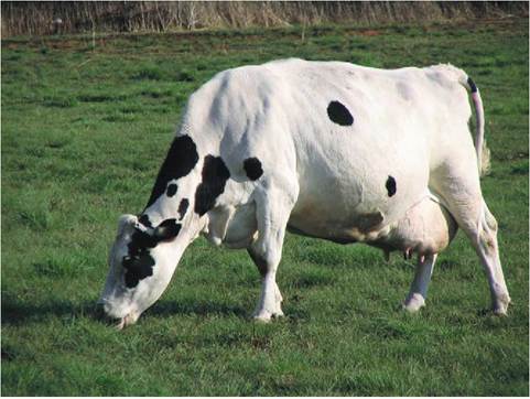
FIGURE 2-21 Organic versus inorganic matter. The molecules that make up a living organism, such as this Holstein cow and the grass she is eating, are primarily organic compounds. Nonliving structures, such as rocks and metal fencing, contain mostly inorganic compounds.
inorganic molecules are essential components of all living organisms.
Why is carbon an essential component of organic molecules? Carbon is small in size and is electrically neutral so it never gains or loses electrons. Instead, it shares electrons with other atoms. Because it has four valence shell electrons, carbon is able to form four covalent bonds with other elements, itnhcelruding o carbon atoms. In this way, carbon enables the formation of long hydrocarbon chains, which can form a linear shape or a ring shape. Collections of atoms called functional groups may be attached to the carbon chains (or rings) and determine the functionality of the molecule as a whole (Figure 2-22). The functional group, though small relative to the entire molecule, is the reactive part of the molecule and determines the molecule's chemical activity.
TEST YOURSELF 2-4
1. What is the difference between organic and inorganic compounds?
2. Are only organic compounds necessary for life?
3. List four types of inorganic molecule that are important for life.
4. List four types of organic molecule that are important for life.
5. What features does carbon possess that makes it particularly well suited for creating the chemistry of living creatures.
6. Define the term "functional group."
INORGANIC COMPOUNDS
WATER
Water is a very simple molecule that has unique properties. As mentioned, the water molecule is composed of one
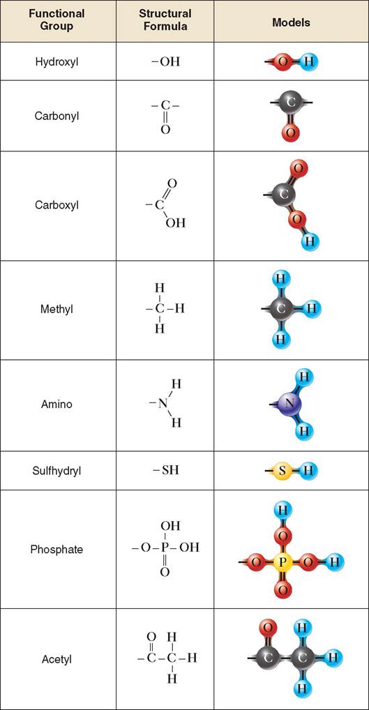
FIGURE 2-22 Common functional groups. Functional groups are attached to the carbon backbone of organic molecules and represent the metabolically active portion of the molecule that determines its biochemical activity.
oxygen atom covalently bonded to two hydrogen atoms. It is a polar molecule that has a slight positive charge in the area of the hydrogen atoms and a slight negative charge in the area of the oxygen atom. This polarity allows water molecules to form hydrogen bonds with each other and with other polar molecules. The polarity of the water molecule allows it to fulfill the following important roles within the living organism.
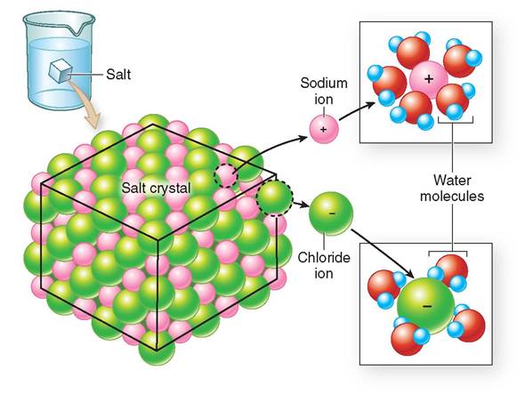
FIGURE 2-23 Water as the universal solvent. A crystal of sodium chloride (NaCl) dissociates in water forming sodium (Na+) and Chloride (Cl-) ions. During this process, water molecules surround each ion. The positively charged ends of the water molecule (hydrogen) are attracted to the negative chloride ions (Cl-), forming hydrogen bonds. Similarly, the negatively charged end of the water molecule (oxygen) is attracted to the positive Na+ ions. In this way, water serves as a universal solvent.

Water is the universal solvent. Chemicals added to water are called solutes, and the combination of the chemicals plus the water is called a solution. More chemicals can be dissolved in water than any other known solvent. Water molecules surround molecules of solute; the negatively charged ends of water surround positively charged molecules, forming a layer of water around each molecule (Figure 2-23). Or, conversely, the positive ends of the water molecule surround or blanket negatively charged molecules. Chemicals that dissolve or mix well in water are called hydrophilic (literally water loving) and are usually polar molecules or ions. Molecules that do not mix well with water are called hydrophobic (water hating). They are usually electrically neutral, nonpolar molecules such as lipids. Hydrophobic molecules gather together into a droplet when they are added to water, and a layer of water molecules then surrounds the droplets. There are no bonds between the water molecules and the hydrophobic molecules (Figure 2-24).
Water is an ideal transport medium. The blanketing property of water allows molecules in the water to move around freely and to be cushioned from each other. Because many molecules dissolve readily in water, they can be carried easily to locations in the body or a cell via blood, lymph, and intracellular and extracellular fluid. Blood is a suspension of cells and chemicals in water and is used to carry cells and chemicals around the body. Urine is a solution of waste products in water and is used to eliminate such waste products from the body.
Water has a high heat capacity and a high heat of vaporization. As the chemicals in solution react, they often give off
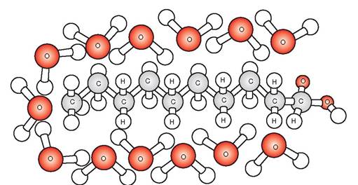
FIGURE 2-24 Water and hydrophobic molecules. Water molecules surround a fatty acid molecule (hydrocarbon chain), which is hydrophobic. No bonds form between hydrophobic molecules and water.
energy as heat. Water is able to absorb heat from biochemical reactions so that the overall temperature of the solution does not rise too rapidly. This stabilization of heat is necessary to keep living organisms in a stable temperature range so that the reactions of life's processes can occur at a steady rate without interruption. The high heat of vaporization means that water needs a fairly high temperature to change from a liquid to a gas; therefore, water will remain in a liquid state through a wide range of temperatures.
Water is used for lubrication. The ability of water to surround molecules allows it to be a lubricant for moving parts in the body. Examples abound in the animal body: the fluid in the pericardial sac allows the heart to move freely within the sac and synovial fluid in the joints allows bones to rub without pain when a limb is moved.
TEST YOURSELF 2-5
1. Why is water called a polar molecule?
2. What are the four properties of water that make it so necessary for life?
SALTS
Salts are mineral compounds that have ionic bonds, and they are the principal form of minerals that enter and are stored in the body. An example of a salt is sodium chloride (NaCl), which is present in large amounts in blood and other tissues. Another example is calcium phosphate Ca3(PO4)2, which is the substance that gives bones their rigidity. When salts are added to water they immediately ionize, or divide into separate ions. Salts in their ionic form are known as electrolytes, substances that have the ability to transmit an electrical charge. The transmission of nerve impulses, for example, requires sodium ions (Na+) and potassium ions (K+). In addition, the contraction of muscle requires sodium, potassium, and calcium ions (Ca2+).

TEST YOURSELF 2-6
1. How is an ion different from an atom?
2. What is an electrolyte?
3. What are some examples of electrolytes?
ACIDS AND BASES
Acids are ionically bonded substances that, when added to water, freely release hydrogen ions (H+). In other words, acids ionize in water and one of their ions is H+. Acids are therefore called H+ donors or proton donors, since H+ is a proton with
no electron. Bases, which are alkaline compounds that are ionically bonded, also ionize in water but release a hydroxyl ion (OH-), not hydrogen ions, therefore bases are known as proton acceptors. Hydroxyl ions are attracted to H+ ions to form water.
Acids and bases are also electrolytes because, when they ionize in water, they can transmit electricity. An example of an acid is hydrochloric acid (HCl). When added to water, the H+ and Cl- ions disassociate and are free to join with other substances. If hydrochloric acid is added to water containing a base such as sodium hydroxide (NaOH), the acid and base neutralize each other. The protons from the acid join with hydroxyl groups from the base and the resulting solution has a neutral pH.

THE pH SCALE
Acidity and alkalinity are measured on a pH scale. The scale ranges from 1 (the most acidic) to 14 (the most alkaline or basic). A pH of 7 is the middle of the scale and is neutral. For example, lemon juice is very acidic and has a pH of 2.3, and ammonia is very basic and has a pH of 11.6. To function properly the tissues and blood in the animal body must maintain a pH of around 7.4, which is slightly basic (Figure 2-25).
BUFFERS
Weak acids and bases are those that do not completely ionize in water. They are important in the physiology of living systems because they act to buffer the solution where chemical reactions take place. Buffering the solution means keeping the pH in the neutral range. As metabolic chemical reactions
∕j CLINICAL APPLICATION
Kidney Failure: Low Potassium
A 16-year-old cat has kidney failure. One of the findings on the serum chemistry test is a low serum potassium concentration. Potassium (K) is an electrolyte that is important for muscle contraction and nerve function. The low level of potassium is caused by the cat’s lack of appetite over the last few weeks: kidney disease makes the cat feel nauseated, so she has a reduced intake of potassium-containing foods and loss of potassium through the damaged kidneys. The lack of potassium makes her feel weak, and it can slow her gastrointestinal contractions and cause constipation. More dangerously, because potassium plays an important role in muscle contraction, the low potassium makes it difficult for the heart muscle to contract. This reduces blood flow to the tissues and can also cause irregular heartbeat; these arrhythmias can be life threatening. We treat this condition by supplementing her intravenous fluids with potassium or by giving her oral potassium supplements.
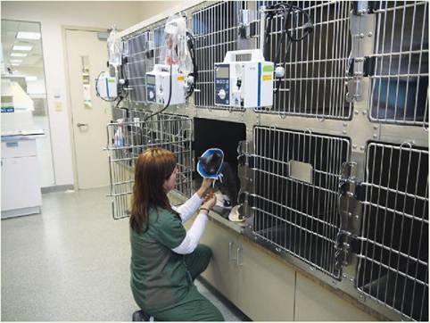
A hospitalized cat in renal failure is examined at regular intervals by veterinary technicians. Fluid therapy is a critical part of treatment. Therefore, an Elizabethan collar is applied to prevent the patient from removing the intravenous catheter. (Courtesy Dr. Joanna Bassert.)
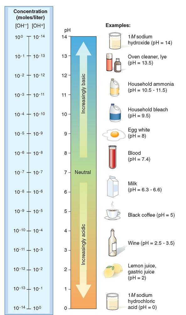
FIGURE 2-25 The pH scale. Many common chemicals and household products are either acidic or basic. As the concentration of H+ increases, the solution becomes more acidic and the pH value decreases. As the OH- concentration increases, the solution becomes more basic or alkaline, and the pH value increases.
take place in the body, strong acids such as lactic acid and strong bases such as ammonia are produced as waste products. If these substances were allowed to accumulate, the pH of the cell or tissue would quickly become too high or too low for chemical reactions to continue. When placed in water, a weak acid will not ionize completely. The product of ionization is a weak base; in other words, a weak acid will initially ionize into free H+ ions, a weak base product, and weak acid molecules (Figure 2-26). The pH of the solution is not changed greatly because some of the chemical remains in acid form and some remains a weak base. If a strong base is added to the solution, the hydrogen ion will attach to the base and neutralize it, and the remaining weak acid will ionize Further.
Buffers help the cell maintain a neutral pH by not allowing excessive hydrogen or hydroxyl ions to accumulate. An example of the most common buffer system is carbonic acid and bicarbonate. Carbonic acid (H2CO3) ionizes, when placed in water, to free hydrogen ions and the weak base, bicarbonate (HCO3-).
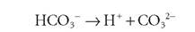
H2CO3 → H++ HCO3-
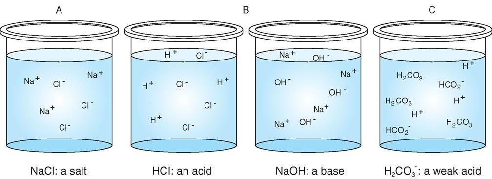
FIGURE 2-26 Salts, acids, and bases. A, When placed in water, salt (NaCl) ionizes completely. B, Hydrochloric acid (HCl) is a strong acid, and sodium hydroxide (NaOH) is a strong base; they ionize completely in water. C, Carbonic acid (H2CO3) is a weak acid and ionizes incompletely to hydrogen ions (H+) and a weak base, bicarbonate (HCO3-).
Bicarbonate (HCO3 ) can further ionize by losing a proton, resulting in carbonate (CO32-).
This means bicarbonate can act as both a weak acid, by losing a proton, and a weak base, by gaining one; this is what makes it such an effective buffer. The complete reaction is written as:
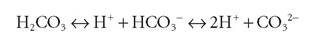
Notice that this equation can run in either direction. What happens when a stronger acid, say lactic acid, is added to the solution? ⅞e strong acid disassociates, and the extra H+ ions increase the speed of the reaction, so more carbonic acid is produced. This is eventually excreted as CO2 via the respiratory system.
H2CO3 + C3H5O3- (lactate) → HCO3- + C3H6O3 (lactic acid)
If a base is added to the blood, the bicarbonate will donate iytdsrhogen ion and more carbonate will be formed.
test yourself 2-7
1. Whichtypeof compound is Icnownasaproton donor: acid or base?
2.What deaspH measure?
i. Is a eolution withte pHofC.d aeidiborbasic?
4.How does a wpaCacldact ara buffer?
∕j CLINICAL APPLICATION
Metabolic Acidosis
Sometimes so much acid accumulates in the animal body that the buffering system is overwhelmed, and the pH of the blood is lowered. This condition is called metabolic acidosis. Two common causes are fatty acid accumulation in diabetes mellitus, ⅛e to the excessive breakdown of lipids for energy, and the accamillation of hydrogen ions in kidney disease caused by the kidney’s inability to excrete them. In these conditions the blood and tissue have a high level of acid: weak acids furfofemrintghe b system and strong acids caused by the
underlying condition. Metabolic acidosis can cause several uncomfortable and dangerous symptoms including anorexia, tvhoamrgityi,ng, le and muscle wasting. Severe metabolic acidosis can decrease cardiac output, reducing blood flow to the thiiscshues, w further damages the kidneys and other organs. Life-threatening cardiac arrhythmias can also develop. Administering balanced electrolyte solutions treats metabolic acido- seis sinc these fluids contain the buffers needed by the body teocrdease the acid concentration in the blood.

A critical canine patient with acidosis receives intravenous fluids with buffers and electrolytes. Humidified nasal oxygen via a nebulizer is also provided to assist with respiration. (Courtesy Dr. Joanna Bassert.)
ORGANIC COMPOUNDS
Organic compounds are composed of large molecules containing carbon. The molecules are divided into four groups: carbohydrates, lipids, proteins, and nucleic acids. What is it about the element carbon that makes it so omnipresent in organic chemistry? Carbon has four electrons in its outer electron shell, so it is moved to share these electrons with other atoms to complete its outer shell. For this reason, carbon is most stable when it has four covalent bonds with other atoms. This allows molecules containing carbon to exist in many forms including chains, rings, and branches. This flexibility allows for various structures to be built using a small selection of atoms. Many of the organic molecules used in the body are macromolecules—long, complex molecules, often with repeating units. Table 2-3 lists the important organic molecules and macromolecules used by the animal body.
Carbohydrates
Carbohydrate molecules are used for energy, storage of energy, and cellular structures; table sugar, starch, and cellulose are all examples. Carbohydrates are composed of atoms of carbon, hydrogen, and oxygen, with hydrogen and oxygen in the same ratio as in water—two to one. You can think of carbohydrates as hydrated carbon or water-containing carbon.
The simplest form of a carbohydrate is called a simple sugar or monosaccharide. Monosaccharides contain three to seven carbon atoms in a chain or ring. An example is glucose, with the chemical formula C6Hi2O6 (Figure 2-27); this molecule is the primary fuel of the body. Since glucose contains six carbon atoms, it is known as a hexose sugar. A sugar with five carbons is a pentose sugar. Another example of a hexose sugar is fructose. It also has the molecular formula C6Hi2O6, but the arrangement of the atoms is different. This molecule is consumed as the primary sugar in fruit and then converted to glucose in the body. Figure 2-28 shows diagrams of these important monosaccharides.
When two monosaccharides are joined together the reaction is a synthesis reaction, and a disaccharide is formed. Because water is created during the reaction—it is extracted from the saccharides—the reaction is called dehydration synthesis. An example is the combination of glucose and fructose to make sucrose, which is table sugar. Cells use synthesis reactions to build molecules needed for cellular functioning. This process is called anabolism. The opposite reaction is when sucrose is decomposed into its monosaccharide components, glucose and fructose. Since water is used in the reaction to break down sucrose this type of reaction is called hydrolysis (Figure 2-29). Cells use decomposition reactions to release energy held in bonds between atoms and to generate the simple molecular building blocks needed by the cell. The decomposition of nutrients is a process called
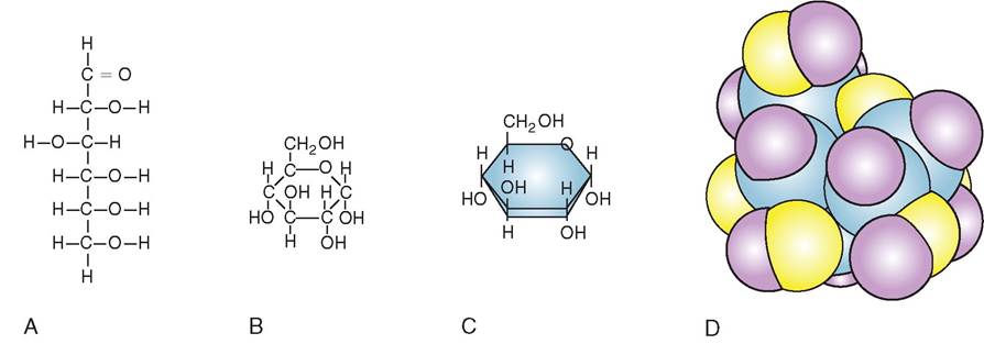
FIGURE 2-27 Structure of carbohydrate molecule. A, The carbon backbone of glucose may form a straight chain. B, A more stable carbon ring structure. C, Shorthand for the glucose molecule, omitting the carbon atoms at the angles of the carbohydrate ring. D, The three-dimensional view of the glucose molecule.
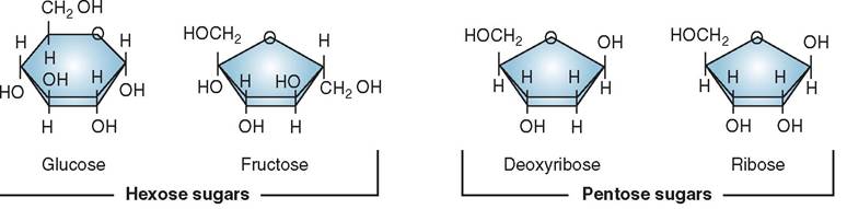
FIGURE 2-28 Monosaccharides. Glucose and fructose are example of hexose sugars. They contain six carbons each. Deoxyribose and ribose are examples of five-carbon pentose sugars.
| TABLE 2-3 Organic Molecules in the Body | ||
| MOLECULAR CLASSIFICATION | CONSTITUENTS AND MOLECULAR STRUCTURE | EXAMPLES AND NOTATIONS |
| Carbohydrates | ||
| Simple carbohydrates | Monosaccharide: most stable as a single pentose ring Disaccharide: two pentose rings | Glucose, fructose, ribose, deoxyribose Sucrose |
| Complex carbohydrates | Polysaccharide | Starches; glycogen: stores energy in liver; cellulose: derived from plants and provides insoluble fiber in diet |
| Proteins | ||
| Amino acids link to form peptide and polypeptide chains; proteins form primary, secondary, tertiary and quarternary structures | Contractile structural proteins that make up muscle (e.g. actin and myosin) Other structural proteins that make up cartilage and tendons (collagen), and hair and skin (keratin) Globular functional proteins such as enzymes, antibodies, hemoglobin and many integral and peripheral proteins in the cell membrane | |
| Lipids | ||
| Neutral fats | Triglycerides: one glycerol molecule (backbone) and three fatty acid chains; fatty acid chains that lack double bonds are "saturated" while those with double bonds are "unsaturated" | Saturated fatty acids are solid (fats) at room temperature and unsaturated fatty acids are liquid (oils) at room temperature; both are concentrated sources of energy |
| Phospholipids | Phosphate head, glycerol backbone and two fatty acid chains | Key component of the bilayer of the cell membrane |
| Steroids | Four flat interlocking rings | Cholesterol, cortisol, testosterone, estrogen |
| Other Lipid Substances | ||
| Fat-soluble vitamins | Variable molecular structure depending upon the specific vitamin; stored in liver and fat | Vitamins A, D, E, and K: stored in body fat; can be toxic if given in excess |
| Eicosanoids | Derived from arachidonic acid, a 20-carbon fatty acid | Regulatory molecules that enhance the immune system and elicit inflammatory responses (e.g., prostaglandins, mediate inflammation; leukotrienes, mediate bronchoconstriction; thromboxanes, mediate platelet function) |
| Nucleic Acids | ||
| Deoxyribonucleic acid (DNA) | Two parallel strands of nucleotides connected via hydrogen bonds; each nucleotide is composed of: a phosphate group, a 2-deoxyribose sugar, and a nitrogenous base (e.g., cytosine, guanine, alanine, or thymine) | Found in the nucleus where it condenses with histone proteins to form chromosomes; also found in mitochondria, providing the molecular instructions for making the enzymes needed for cellular respiration |
| Ribonucleic acid (RNA) | A single strand of nucleotides; each nucleotide is composed of: a phosphate group, a ribose sugar, and one nitrogenous base (e.g., cytosine, guanine, alanine, or uracil) | Three types of RNA: ribosomal RNA (rRNA), forms ribosomes and found in the nucleolus; transfer RNA (tRNA), carries amino acids to docking stations on mRNA; messenger RNA (mRNA), carries the genetic code from the nucleus to the cytosol |
| Adenosine triphosphate (ATP) | An ATP molecule is composed of: adenine, a ribose sugar, and three phosphate groups | ATP is used by the cell to carry out all active metabolic processes; when the terminal phosphate group is removed, energy is released and the molecule becomes adenosine diphosphate (ADP). ADP is transported back to the mitochondria where it is converted back to ATP |
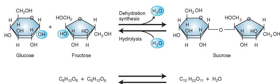
FIGURE 2-29 Disaccharide. Glucose and fructose are joined in dehydration synthesis to make sucrose, a disaccharide, and water. The opposite reaction, hydrolysis, decomposes sucrose into glucose and fructose.
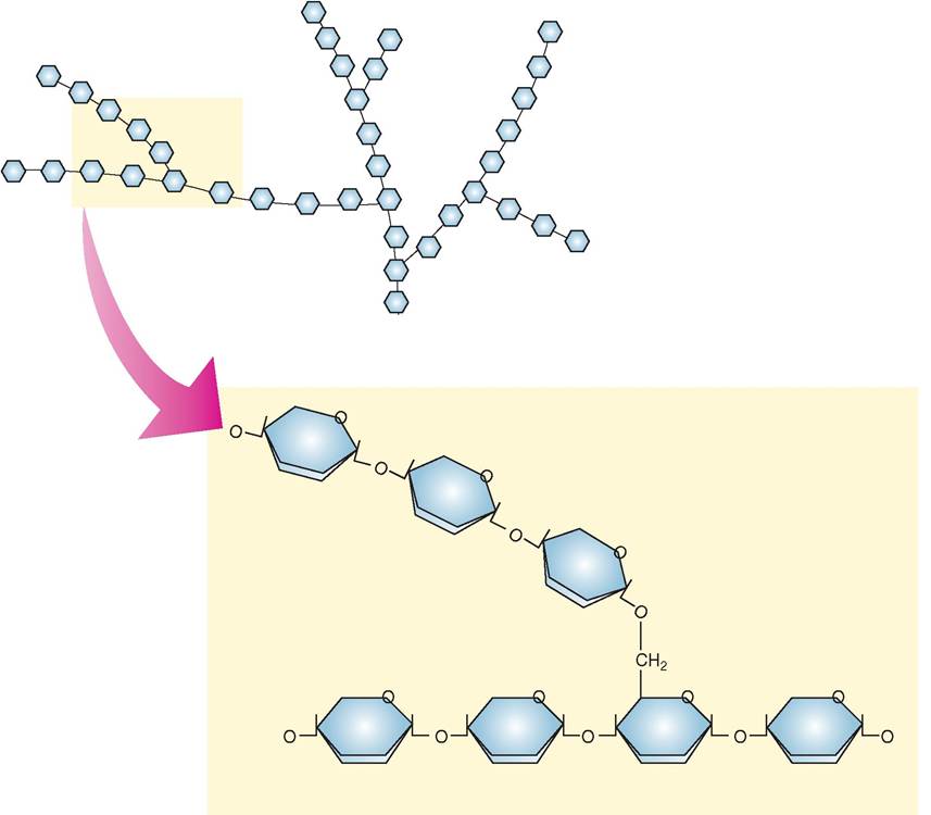
FIGURE 2-30 Polysaccharide. A simplified model of a molecule of glycogen. Glycogen is a polysaccharide made of the multiple glucose molecules bonded in a branching chain. Glucose is stored in the liver in form of glycogen. The branching of the molecules creates many ends that allow for reactions at each end to break glucose molecules off very rapidly when the animal needs energy.
catabolism, and this process, along with anabolism, will be explored in more detail in Chapter 17.
Polysaccharides are combinations of many monosaccharides, all joined by dehydration synthesis. Polysaccharides can have a structural or a fuel storage function. Glycogen (Figure 2-30) is an important polysaccharide that stores fuel in body tissues, and starch is a polysaccharide that has a similar function in plant tissues. Cellulose is the most abundant organic molecule in the biosphere; it is a polysaccharide that provides structural strength to plants. Herbivores can digest cellulose and use the component monosaccharides as fuel.
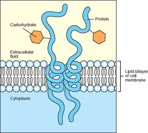
FIGURE 2-31 Glycoprotein. Glycoproteins, macromolecules made of amino acid and carbohydrate units, are found in cell membranes. The carbohydrate recognizes molecules that are to be transported into the cell by the protein channel.
Carbohydrates can also be joined to other molecules, such as proteins or lipids, to create macromolecules important to life. For example, a glycoprotein is a macromolecule composed of a carbohydrate attached to a protein. The carbohydrate component of a cell membrane glycoprotein has important roles in the adhesion of the cell to other cells and in recognition of molecules to be transported into the cell (Figure 2-31).
LIPIDS
Lipids are used in the body for energy and are stored in fat for future energy needs. Lipids serve as chemical messengers in the form of some hormones. There are four classes of lipid that are important for life: neutral fats, phospholipids, steroids, and eicosanoids. Like carbohydrates, lipids are made of carbon, hydrogen, and oxygen, though their oxygen content is much lower. They also sometimes contain phosphorus.
NEUTRAL FATS
Neutral fats are also called triglycerides or, simply, fats. A triglyceride contains three fatty acids (hence the “tri” in the name) and a glycerol molecule. A glycerol molecule is a modified, three-carbon simple sugar. It has the formula C3H8O3. A fatty acid is a chain of carbon atoms with one or two hydrogen atoms attached to each carbon by single or double bonds. A fatty acid is called a saturated fatty acid when all the bonds in the hydrocarbon chain are single bonds and as many hydrogen atoms as possible are attached to the carbon. Saturated fatty acids are mainly found in animal fats such as butter and lard. A fatty acid is called an unsaturated fatty acid when there are some double bonds between the carbon and hydrogen atoms. Unsaturated fatty acids are mainly of plant origin, such as corn oil and olive oil. The glycerol molecule and the three fatty acid molecules connect together in the shape of an “E” with the glycerol molecule making the backbone of the E (Figure 2-32). They are joined by dehydration synthesis: three water molecules are produced by joining the hydrogen from the fatty acids to the hydroxyl (OH) groups of the glycerol.
TEST YOURSELF 2-8
1. What three elements are found in all carbohydrates?
2. What is the name of a simple sugar?
3. What process joins multiple simple sugars?
4. What is another name for a complex, multiunit carbohydrate?
When triglycerides are decomposed, the reaction is called hydrolysis, and water molecules are consumed. These reactions should be familiar because they are similar to the reactions that occur with carbohydrate synthesis and decomposition. Neutral fats are mainly used for energy; the body gets energy by breaking down the bonds in the neutral fats and stores energy by transporting the excess neutral fats to adipose tissue. The fat-filled cells act to pad vital organs from trauma and act as insulation to help maintain body temperature. Neutral fats are hydrophobic and do not mix in water.
A lipoprotein is a macromolecule composed of proteins and lipids. Lipoproteins are used to transport fats within the body. The hydrophilic proteins allow the fats to be shielded from the blood plasma and to be transported.
PHOSPHOLIPIDS
Phospholipids are similar to triglycerides in that they have a glycerol backbone. They have two fatty acids attached to the glycerol extending in one direction. In place of the third fatty acid, they have a phosphate group (PO4) attached to a nitrogen-containing compound extending in the other direction. The phosphate group side, or head end of the phospholipid, is water soluble, meaning it is hydrophilic and polar, whereas the fatty acid side, or tail end of the molecule, is water insoluble, that is, hydrophobic and nonpolar (Figure 2-33). This unique property is what makes phospholipids line up in two layers, called a lipid bilayer, when placed in a polar substance such as water. The hydrophilic heads form hydrogen bonds with the water and the tails are repelled from the water and are most stable when abutting another tail. Phospholipids are the main component of cellular membranes. They also form the myelin sheath of nerve cells.
STEROIDS
Steroids are lipids that take the form of four interlocking hydrocarbon rings. They are hydrophobic, nonpolar substances with very little oxygen. Different types of steroid are formed by attaching unique functional groups to the four- ring structure of the molecule (Figure 2-34). The basic cholesterol ring structure is synthesized from acetyl CoA (see
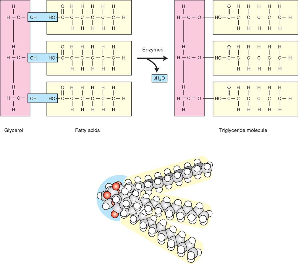
FIGURE 2-32 Triglyceride. A triglyceride molecule is formed by dehydration synthesis and is composed of three fatty acids and one glycerol molecule. A three-dimensional view is also shown.
Chapter 17). Cholesterol is used in the formation of bile salts, which aid in fat digestion. Cholesterol is also used by the adrenal glands, testes, and ovaries for creation of steroid hormones including cortisone, estrogen, progesterone, and testosterone.
EICOSANOIDS
Eicosanoids are lipids formed from a 20-carbon fatty acid and a ring structure (Figure 2-35). The prefix eicosa derives from the Greek word for twenty. Eicosanoids are important substances for the mediation of complex chemical processes in the body and include: prostaglandins (PGs), which mediate inflammation; thromboxane, which mediates platelet function; and leukotrienes, which mediate bronchoconstriction and increased mucus production.
TEST YOURSELF 2-9
1. What three elements are found in all lipids?
2. Which atom makes up the backbone of all lipid molecules?
3. Which lipids are polar: neutral fats or phospholipids?
4. What is the function of lipids in the body?
PROTEINS
Proteins are the most abundant organic molecules in the body. They also have the widest variety of functions (Table 2-4). Proteins are used for cell structures and structural body tissues, for controlling chemical reactions, for regulating growth, and for defending the body from invaders. Proteins catalyze or speed up all reactions occurring in the body, and
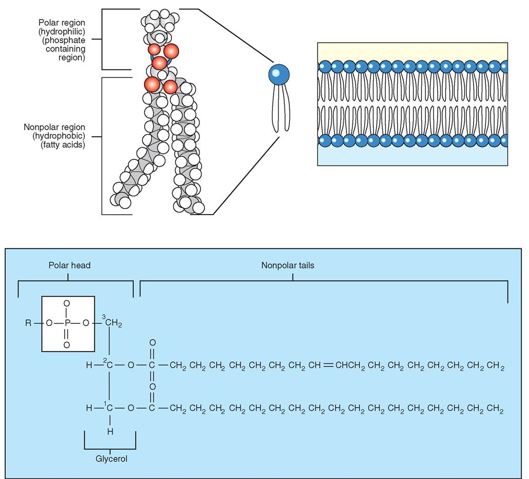
FIGURE 2-33 Anatomy of a phospholipid. Example of hydrophilic and hydrophobic regions of a phospholipid molecule. In the diagram of the cell membrane, note that the phospholipids form a lipid bilayer with the polar part of the phospholipid molecules hydrogen-bonded to and facing the water molecules.
| TABLE 2-4 Functions of Proteins | ||
| PROTEIN STRUCTURE | FUNCTION | EXAMPLE |
| Functional (globular) | Chemical reactions | Protein enzymes: essential to almost every biochemical reaction in the body |
| Transport of molecules | Hemoglobin transports oxygen in the blood | |
| Regulation of metabolism | Peptide hormones: regulate metabolic activity, growth, and development (e.g., thyroid hormone regulates metabolic rate and insulin regulates blood sugar levels) | |
| Immune system | Antibodies (immunoglobulins) are proteins created by immune cells that recognize foreign substances such as viruses | |
| Structural (fibrous) | Structural framework | Collagen: gives strength to bones, tendons, ligaments Keratin: hair, nails, waterproofing of skin |
| Physical movement | Actin and myosin: contractile proteins found in muscle; actin also used for intracellular transport | |
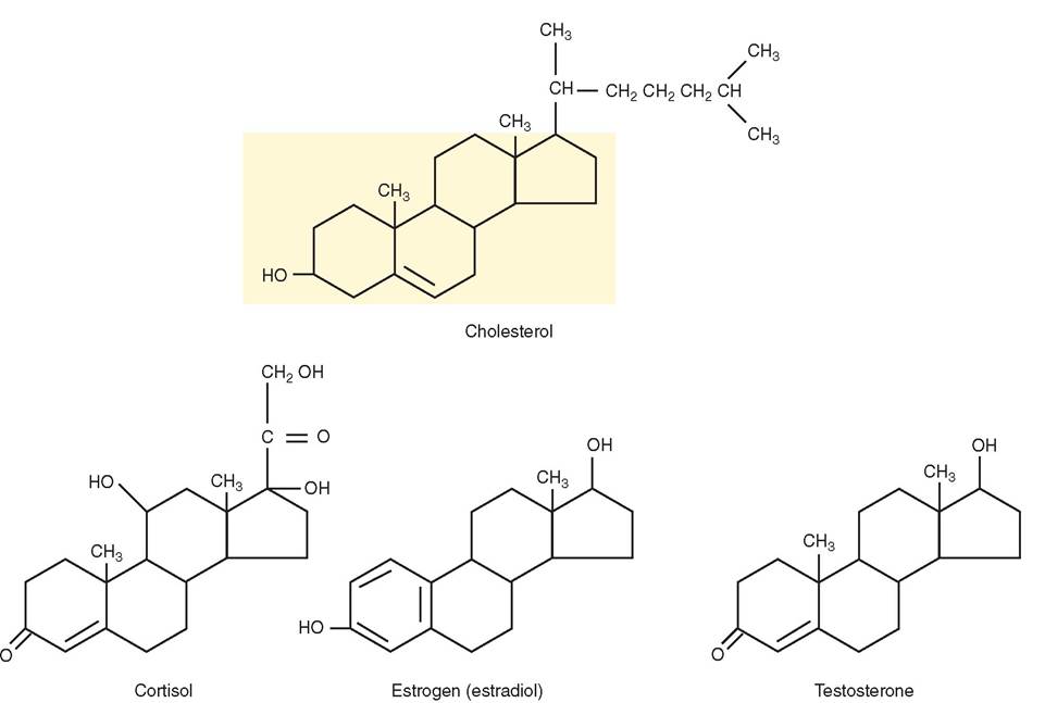
FIGURE 2-34 The steroid nucleus. The steroid nucleus (highlighted) found in cholesterol is a four-ring structure. Attaching a different functional group to the basic four-ring structure forms a different steroid compound. You can see how easy it is for any steroid to be converted into another type of steroid. The clinical significance of this is exemplified by the high cholesterol seen in horses and dogs with Cushing's syndrome (high cortisol is converted to cholesterol). This also plays an important role in women with breast cancer. Hormones in meats, dairy products, and some therapeutic drugs consumed by women, as well as Xenoestrogens found in insecticides and other environmental pollutants, can be converted to estrogens and other tumor-enhancing steroid hormones (progesterone).
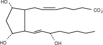
FIGURE 2-35 Eicosanoid. This is a diagram of prostaglandin F2alpha, which is used to lyse the corpus luteum to alter the reproductive cycle in female cattle. The 20-carbon fatty acid is in the classic hairpin formation of all eicosanoids.
they transport ions and other molecules into and out of the cell and around the body. You can think of proteins as the worker molecules of the body that organize and facilitate all metabolic processes. Proteins are organic molecules made chiefly of carbon, oxygen, hydrogen, and nitrogen, though some proteins also contain sulfur, iron, or phosphorus. The building blocks of proteins are amino acids, linked together like the cars of a long train. The sequence of the amino acids is what makes each protein unique and defines the function of the protein.
AMINO ACIDS
There are 20 different amino acids used by the body, but they all share the same basic structure. The amino acid molecule contains a central carbon atom attached to a hydrogen atom, an amino group (NH2), a carboxyl group (COOH), and a unique group of atoms called a side chain designated by the letter R (Figure 2-36). The R group defines each amino acid. Amino acids can be linked together in an infinite variety of combinations to form proteins. The specific combination of amino acids is ordered by the cell's DNA, and this is what determines the nature and function of the resultant protein. Two amino acids are linked together by a dehydration synthesis reaction. The carboxyl group of one amino acid links with the amino group of another amino acid via a peptide bond. A short chain of two amino acids is called a dipeptide (Figure 2-37). A tripeptide is a chain of three amino acids linked together, and a polypeptide is a chain of 10 or more amino acids linked together. When the chain exceeds 100 amino acids it is called a protein.
STRUCTURE OF PROTEINS
The shape of a protein molecule directly determines its function. For example, fibrous proteins such as collagen are
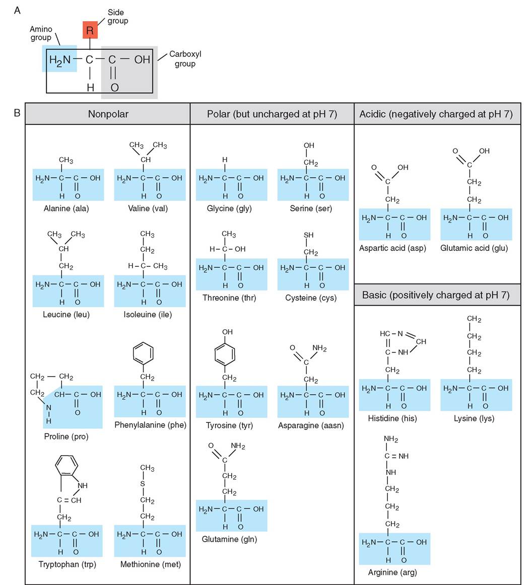
FIGURE 2-36 Structure of amino acids. A, Every amino acid is composed of an amino group, carboxyl group, and side chain (R). The side group can be simple or complex, depending on the amino acid. B, The 20 standard amino acids required for animal life to exist. Alanine is an example of an amino acid with a simple side group. Tryptophan has a more complex side group.
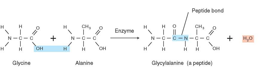
FIGURE 2-37 Formation of a dipeptide. A dipeptide is formed when two amino acids combine by dehydration synthesis. The carboxyl group of one amino acid is bonded with the amino group of another amino acid by a peptide bond, releasing a molecule of water.
long and firm, which allows them to be used to add strength to tissues, for example the connective tissue in ligaments and tendons. Globular proteins such as immunoglobulins, also known as antibodies, have a specific shape, so they may join like a puzzle piece with a specific foreign protein that invades the body. The structure of proteins is often described in four levels. The primary structure is the sequence and number of amino acids that link together to form the peptide chain. The secondary structure is the natural bend of parts of the peptide chain as it is formed in three dimensions. The bends are stabilized when the atoms of the protein molecule form weak hydrogen bonds with each other (Figure 2-38).
The most common shapes that chains of amino acids assume are the alpha helix and the beta-pleated sheet. The alpha helix occurs when the chain of peptides winds into a spring shape, like a “Slinky” toy or a curl of hair. A betapleated sheet occurs when the peptide chain folds into a shape like an accordion. These shapes can both occur within the same protein at different places in the peptide chain. The tertiary structure is the overall shape of a single protein molecule. A protein molecule further folds in on itself, often shielding the inner, hydrophobic amino acids from the watery environment of the cell or blood. The outer, hydrophilic amino acids allow the protein to be water soluble. The folds are held in place by more hydrogen bonds and also some covalent bonds, such as the disulfide bond, which is a sulfur atom in one part of the protein covalently bonded to a sulfur atom in another part of the protein. The quaternary structure is when two or more protein chains join to form a complex macromolecule. Again hydrogen and covalent bonds between atoms of the proteins stabilize the shape of the macromolecule.
STRUCTURAL PROTEINS
Structural proteins are stable, rigid, water-insoluble proteins that are used for adding strength to tissues or cells. Because they often have a long, stringy shape they are called fibrous proteins. Examples include: collagen, which is the main protein in connective tissues such as ligaments, cartilage, bone, and tendons; fibrin, which is the fibrous connective tissue in blood clots; and keratin, which is the main protein in hair, hooves, horns, and the outer layer of skin.
FUNCTIONAL PROTEINS
Functional proteins are generally water soluble and have a flexible, three-dimensional shape, which can change under different circumstances. Because they have a convoluted, changeable shape they are called globular proteins. Globular proteins are highly chemically active molecules. Examples include hemoglobin, antibodies, protein-based hormones, and enzymes, which are proteins that catalyze or speed up chemical reactions (Figure 2-39).
HOW ENZYMES WORK. Enzymes are essential to the body in their role of catalyzing chemical reactions; without them, most chemical reactions in the body would occur too slowly to produce the chemicals needed. Enzymes speed up a chemical reaction without being destroyed or altered, and they are specific to the reaction that they catalyze and to their substrates, which are the substances they act upon. This specificity is determined by the shape, charge, and hydro- philic/hydrophobic properties of the enzyme and its substrates. This specificity is often referred to as the lock and key property of enzymes, because an enzyme fits its substrates exactly and is itself unaltered at the end of the reaction. Enzymatic reactions often take place in a series of reactions with the products of one reaction acting as the substrate for the next reaction. Often these reactions occur along lipid membranes with the enzymes existing as proteins within the membrane aligned in order of the reaction. An example of this is seen in the description of the Krebs cycle and the electron transport system for the production of ATP molecules (see Chapter 17).
TEST YOURSELF 2-10
1. What element is found in all proteins that is not found in carbohydrates or lipids?
2. What is the building block for proteins?
3. What is the name of the bond holding two amino acids together?
4. What is a peptide?
5. How does an enzyme work?
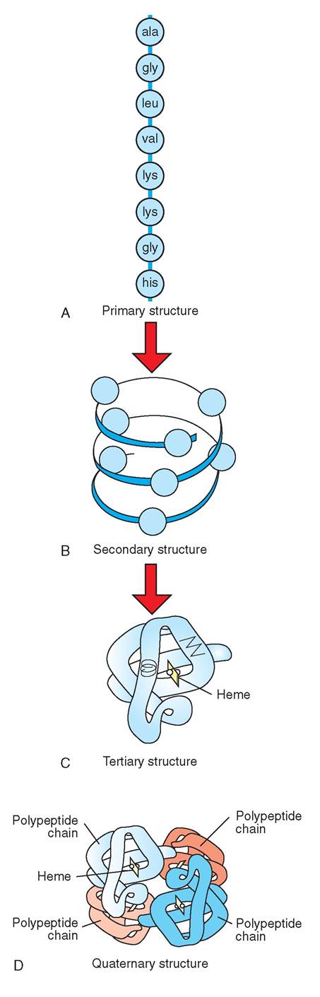
FIGURE 2-38 Levels of organization of proteins. A, The primary structure of proteins consists of a sequence of amino acids. Amino acids are linked to one another like beads in a necklace. B, The secondary structure can be either helical or pleated and is held by hydrogen bonds between nearby amino and carboxyl groups. C, The tertiary structure consists of either folded alpha helixes or beta pleats. D, The quaternary structure refers to the combination of more than one polypeptide chain; such chains unite to form the complete protein molecule.
CLINICAL APPLICATION
Hyperthermia and Protein Denaturation
Hyperthermia is the scientific name for elevated body temperature, and it can have many causes such as a fever, heatstroke, or prolonged seizures. When the body temperature becomes too elevated for too long, the chemical bonds between and within molecules start to break. The hydrogen bonds holding proteins in their tertiary and quaternary structures are especially sensitive to this stress. When these bonds break, proteins are released from their complex structures and stretch into a straight chain of amino acids. Because they no longer have their unique shape these proteins lose their function. This is called the denaturation of proteins. Once this happens on a large scale it is irreversible, and the tissues of the body are irreversibly damaged. Some body proteins denature at 40° C (104° F), and death will usually occur around 41.7° C (107° F) if that temperature is maintained for 30 minutes.
NUCLEIC ACIDS
Nucleic acids are the largest molecules in the body and are composed of carbon, oxygen, hydrogen, nitrogen, and phosphorus. There are only two classes of nucleic acids: DNA and RNA. DNA or deoxyribonucleic acid exists mainly in the nucleus but also in mitochondria; it is the molecule that contains all the instructions needed by the cell to build proteins. These instructions determine the shape and function of every tissue in the body and therefore the shape and function of the living organism (Figure 2-40). The instructions are coded in segments of the DNA called genes. RNA or ribonucleic acid transfers the instructions out of the nucleus and into the cytoplasm of the cell and builds the proteins. You can think of DNA as the blueprint for the cell and the RNA as the scanner/fax/printer that brings the instructions to where they are needed. The proteins created then catalyze all the reactions performed in the cell, creating and breaking down all the substances needed by the body.
NUCLEOTIDES
The molecular building blocks of nucleic acids are the nucleotides. There are five different nucleotides but they all have the same basic structure. They are composed of a nitrogenous base, plus a five-carbon (pentose) sugar, plus a phosphate group (Figure 2-41). The sugar in DNA is
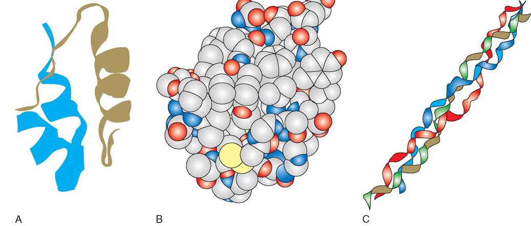
FIGURE 2-39 Exampl es of proteins. A, Proteins are commonly depicted in a ribbon-like form to show the complex secondary and tertiary structure of the chain of amino acids. Here is a model of the insulin molecule showing the helical and straight chains of amino acids. B, A three-dimensional view of the atoms of a functional (globular) protein (insulin) shows the true space-filling, solid shape of proteins. The solid shape is essential to the specific function of the protein because the shape determines which substrates will react with the protein. C, Interwoven strands of amino acid chains are used to create structural (fibrous) proteins. The protein collagen has three strands of amino acids woven in a coil.

FIGURE 2-40 Differences in living organisms. DNA is the nucleic acid that forms genes. Genes determine the shape and function of all living tissues and therefore the shape and function of living organisms. Even in animals with almost completely identical genes, such as within the same species, there are vast differences in how the animals look and how they behave.
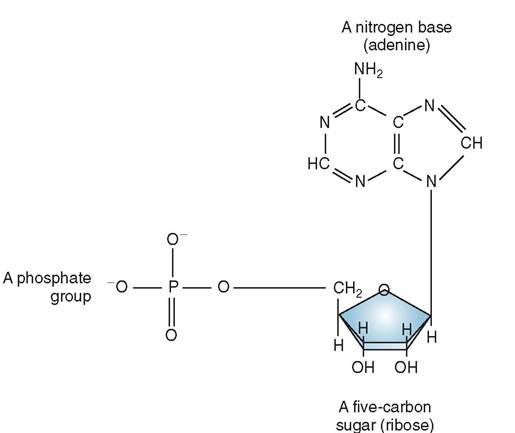
FIGURE 2-41 Nucleotide. Nucleotides are the building blocks of nucleic acids. Each nucleotide contains a phosphate group, a five-carbon sugar, and a nitrogenous base.
deoyxribose and the sugar in RNA is ribose, thus giving DNA and RNA their respective names. The five nucleotides are named for their nitrogenous base: adenine, guanine, cytosine, uracil, and thymine. Three nucleotides, adenine (A), guanine (G), and cytosine (C), occur in both DNA and RNA. Uracil (U) occurs only in RNA and thymine (T) occurs only in DNA.
The nucleic acid is formed by the sugar and phosphate groups joined in a long chain with the nitrogenous base open and available for metabolic activity (Figure 2-42). The information needed to produce proteins is determined by the order of the nucleotides; a grouping of three nucleotides is the code for a specific amino acid. For example, C-G-T is the code for the amino acid alanine. A gene is a sequence of
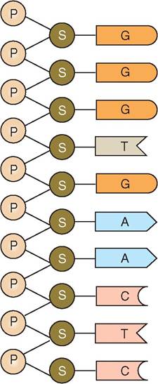
FIGURE 2-42 Segment of a nucleic acid molecule, DNA. The phosphate of one nucleotide bonds with the sugar group of another nucleotide. These phosphate-sugar bonds make up the 'backbone' of the nucleic acid molecule. A nitrogenous base sequence is formed, which provides the cell with coded information. The exposed nitrogenous base is available to bond to a nitrogenous base that is part of a complementary chain.
nucleotides that carries the information to make one peptide chain. Long chains of genes are combined with protein to form chromosomes. Chromosomes replicate during cell division so that all daughter cells inherit an identical copy of the chromosomes from the parent cell.
DNA
Deoxyyribonucleic acid molecules consist of two parallel strands of the nucleotides adenine, guanine, cytosine, and thymine. The strands are connected by hydrogen bonds between the nitrogenous bases. Each nitrogenous base can only bond with one other, specific nitrogenous base: adenine can bond only with thymine and guanine can bond only with cytosine. The two strands of bonded nucleic acid twist around each other in a spiral called a double helix (Figure 2-43). The order of the nucleotides is unique to each individual and is carried in every cell of the individual.
RNA
RNA consists of only one strand of the nucleotides adenine, guanine, cytosine, and uracil. There are three types of RNA that have unique roles in protein synthesis: transfer, messenger, and ribosomal RNA. Transfer RNA copies the information in the DNA molecule, messenger
TEST YOURSELF 2-11
1. How does a nucleotide differ from an amino acid?
2. What three parts compose a nucleotide?
3. How many nitrogenous bases are there?
4. Explain the structure of DNA.
5. Why is DNA important to life?
CLINICAL APPLICATION
Genetic Disease: Be Careful What You Breed For
Genetic disease is caused by inheritable mutations of DNA. Any change to DNA, such as insertion, deletion, or mixing of the nitrogenous base pairs, leads to a nonsense sequence in the DNA. This defect can manifest as a disease in an organ system. Genetic diseases exist that affect the eyes, skeleton, skin, kidney, immune system, and enzymatic pathways. The defect may result in an incidental finding, such as microcytosis in Akitas, or the defect may cause life-threatening organ failure, such as copper-storage disease in Bedlington Terriers.
Owing to the frequency of inbreeding in small animals, the most common type of disease that is seen is caused by inheritance of recessive traits. To inherit a recessive trait the animal must receive an affected chromosome from both parents. Obviously, affected animals are not bred, but a carrier animal may be inadvertently used in the breeding program. A carrier animal appears unaffected because it has an unaffected chromosome that is dominant over the recessive, affected chromosome. If two carrier animals are bred, each of the offspring has a 25% chance of being affected, inheriting the affected chromosome from each parent. They have a 50% chance of being a carrier by inheriting an affected chromosome from only one parent. When choosing which animals to breed, the ethical breeder will not breed relatives of any affected animals. If a relative of an affected animal must be used for breeding, there are tests available for some genetic diseases that will confirm whether or not the animal is a carrier. Some tests measure the production of abnormal proteins generated because of the DNA mutation. Carriers usually produce a small amount of the abnormal proteins. DNA tests can also detect the mutant genes for some diseases directly.
RNA carries the information out of the nucleus, and ribosomal RNA uses the information to create the proteins needed by the body.
ATP
When a cow eats its meal of grain and grasses, the food is broken down into nutrients such as glucose and proteins. Glucose that is derived from food is converted into energy by the cells. As glucose is broken down into monosaccharides, the energy created is stored in a molecule called adenosine triphosphate (ATP). You can think of
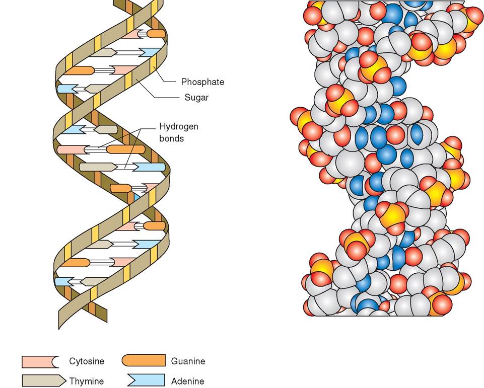
FIGURE 2-43 Structure of the DNA double helix. Deoxyribonucleic acid (DNA) looks like a spiraling ladder, the vertical portion of which is composed of alternating phosphate and sugar groups; the 'rungs' of the ladder are composed of nitrogenous bases. A three-dimensional model is also shown.
ATP as the energy currency of cells. The cells need ATP to fuel any work that they do. Just as wood is added to a fire, and the fire burns the wood to create heat, nutrients are added to the body, and the cells use up the nutrients in a process called cellular respiration to create ATP (see Chapter 17).
The energy needed by the body is stored in the phosphate bonds of the ATP molecule. ATP is an RNA nucleotide containing the nitrogen base adenine with two additional phosphate groups attached (Figure 2-44). The bonds between the phosphate groups are called high-energy bonds. It is when these bonds are broken that energy is released from the ATP molecule. To use the energy stored in ATP, enzymes must move the terminal phosphate group to another molecule. The receiving molecule is then termed phosphorylated and temporarily has energy to do some work. During this process
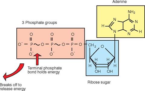
FIGURE 2-44 Structure of ATP. ATP is formed from a ribose sugar, the nitrogenous base adenine, and three phosphate groups. Bonds between the phosphate groups hold energy.
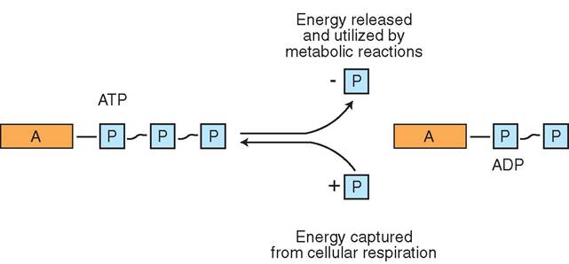
FIGURE 2-45 Conversion of ATP to ADP. Energy is released and available for cellular work when a phosphate group breaks off from the ATP molecule. The resultant molecule, with two phosphate groups, is ADP. A phosphate group can be added back to the ADP, creating ATP through the process of cellular respiration.
the ATP molecule loses a phosphate group and becomes adenosine diphosphate (ADP) (Figure 2-45).
Another phosphate group can be used, resulting in the creation of a molecule of adenosine monophosphate (AMP). As more glucose and other nutrients are metabolized, phosphate groups are joined to AMP, creating a renewed source of ATP. Without ATP, muscle cells could not contract, enzymatic reactions could not take place, and molecules could not be actively transported across cell membranes.
TEST YOURSELF 2-12
1. How does an ATP molecule differ from a nucleotide?
2. How does an ATP molecule supply a cell with energy to do work?