CONTAGIOUS ECTHYMA
TURID VIK0REN
Norwegian Veterinary Institute, Oslo, Norway
Contagious ecthyma (CE) (synonyms: orf, contagious pustular dermatitis) is a viral skin disease caused by the Orf virus or other closely related viruses within the genus Parapoxvirus (Family Poxviridae).
The disease is common among domestic sheep and goats worldwide, and has been diagnosed in several wild species and humans (it is a zoonosis).The distribution and host range among wild artiodac- tyls have been summarized by Robinson & Kerr(61). Among free-living wild species in Europe, CE has been reported in ibex ( Capra ibex), chamois (Rupicapra rupicaprd)6τ, and southern chamois (Rupicapra pyrenaica)es~, in the Alps and Pyrenees, and in a small Norwegian population of musk ox ( Ovibos moschatusfi't. Disease outbreaks have also been reported in semi-domesticated reindeer (Rangifer tarandus) in Fennoscandia(65).
Experimentally, calves of moose (Alces alces), wapiti (Cervus elaphus nelsons), white-tailed deer (Odocoileus vir- ginianus) and other deer species have been shown to be susceptible to CE infection, but the lesions were mild(61). Presumed infection with parapoxvirus has been reported in free-ranging red deer (Cervus elaphus) calves from Germany with the ‘labial form’ of CE(66). A pustular dermatitis caused by a unique parapoxvirus has occurred in farmed red deer in New Zealand, but is not reported from Europe.
The parapoxviruses causing CE in musk ox and reindeer in Norway are genetically similar to Orfvirus isolates from sheep(64’67), whereas the Finnish parapoxvirus isolates from reindeer have been grouped with either Orfvirus or bovine Pseudocowpox virus(68).
Young individuals seem to be more susceptible to CE than adults, and they develop lesions of greater severity; however, older animals are also affected.
Morbidity can approach 100%, but mortality is usually low in uncomplicated cases. Secondary bacterial or fungal infections cause increased mortality1-64’69).Transmission of parapoxvirus is directly by contact with affected animals or indirectly through contact with infectious virus in the environment. Virus is shed in scab material and can survive in dry scabs for months or years in a dry environment sheltered from rainfall(69). The source of infection in free-ranging wild species is usually difficult to identify, but transmission from domestic sheep and goats that graze on the same pastures as wild species is a possibility. Also, subclinical parapoxvirus infections within the wild populations themselves might cause clinical CE outbreaks if triggered by reduced nutritional status, immunosuppression and/or environmental predisposing factors such as stress, handling and fencing. A dense population with high stress levels, in combination with many calves immunologically naive to parapoxvirus, were given as a possible explanation for trigging a severe outbreak of CE among free-ranging musk ox in Norway)64).
Orf virus is epitheliotrophic and infects via damaged skin and replicates in epidermal keratinocytes. The skin infection progresses from erythema to papule, vesicle, pustule and then to scab within a week after infection. Additional pustules and scabs form during the following days. The lesions can be highly proliferative, developing into wart-like papillomas. Primary lesions usually resolve within 4 weeks, usually without scar formation. In uncomplicated cases, CE is febrile and self-limiting)69).
Pustular scabby lesions on the lips and in the skin around the mouth and nostrils are characteristic for CE, but lesions may also develop in the skin or mucosa on other parts of the body such as the buccal cavity (hard palate, tongue, gingiva), limbs (coronet, distal limb), udder, genital skin and oesophagus.
Lesions are seldom found in other internal organs)69). There is variation in the severity of outbreaks and in the anatomical distribution of lesions. In severe cases, large coalescing, papillomatous, cauliflower-like lesions are seen (Figure 13.1). The lesions often ulcerate, and secondary bacterial or fungal infection may occur, often with subsequent lymphadenitis. Infestation with fly larvae (cutaneous myiasis) is also seen. Severe lesions on the mouth/oral mucosa and feet interfere with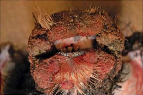
FIGURE 13.1 A musk ox calf from a free- ranging population in Norway suffering from contagious ecthyma. Severe, multiple to coalescing, ulcerated, papillomatous lesions are seen on the lips and the skin around the mouth.
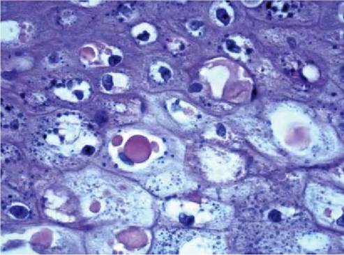
FIGURE 13.2 Microphotograph of a contagious ecthyma lesion in the skin of a musk ox calf showing part of the epidermis. Intracytoplas- mic eosinophilic inclusion bodies are seen in degenerating keratinoc- ytes. Haematoxylin and eosin stain. Magnification: 630?.
normal feeding and locomotion, causing lameness, starvation and loss of body condition)64).
The histopathological picture seen in CE is a pustular dermatitis characterized by epidermal proliferation, degenerating keratinocytes with intracytoplasmic eosinophilic inclusion bodies (Figure 13.2), vesicopustules, microabscesses and multifocal ulcerations in the epidermis which is covered by a serocellular crust)64).
In sheep, there is an Orf virus-specific cellular and humoral immune response after infection, but immunity seems to be short-lived. Thus, the Orfvirus can repeatedly infect sheep and goats; however reinfection lesions are smaller, less severe and resolve more rapidly than primary lesions)70). For further reading regarding Orfvirus infection and host immunity, including viral virulence and immunomodulatory factors the reader may consult Haig )2006))70).
A diagnosis of CE is made by a combination of clinical signs, pathological findings and detection of the agent. Electron microscopy of tissue samples from CE lesions shows typical parapoxvirus particles and is a reliable method if sufficient intact virus particles are present. PCR methods and sequencing of viral DNA are now becoming the tool of choice for detection of the virus.
A disease outbreak might contribute to a transient population reduction and a temporary change in the age distribution within the population, as seen in a free-ranging musk ox population in Norway. The long-term impact on the population, however, appeared negligible)64). Severely affected wild animals should be euthanized for animal welfare reasons.
More on the topic CONTAGIOUS ECTHYMA:
- Tuberculosis in humans and animals is an ancient contagious disease, with a worldwide distribution.
- 5 Appendices