Control of respiration: neural mechanisms
The medullary rhythmicity area is located in the medullar oblongata, and it controls the basic respiration rhythm. It consists of two areas, the inspiratory
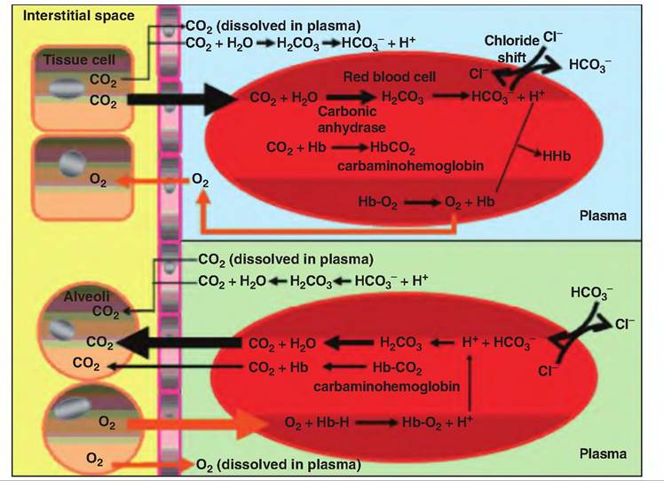
Fig.
14.11. Transport of O2 and CO2 in the blood. The top portion of the figure shows gas exchange at the tissue level, and the bottom portion is at the level of the lungs. As CO2 is produced in the tissues, it diffuses into the red blood cells (RBCs) where it combines with water to form carbonic acid. As carbonic acid dissociates, the HCO3- that was formed diffuses out of the RBC and into the plasma in exchange for Cl", which moves into the RBC, a process called the chloride shift. Most CO2 (70%) is transported as HCO3-; the remainder is transported as either dissolved CO2 (7%) or Carbaminohemoglobin (22%). The bottom portion of the figure shows how the gases move when the blood arrives in the lungs. Hb, hemoglobin.and expiratory areas, also called the dorsal respiratory group and the ventral respiratory group, respectively. The inspiratory area sends signals to the diaphragm via the phrenic nerves and to the external intercostal muscles via the intercostal nerves. These signals cause muscle contraction resulting in inspirations. When these signals cease, inspiration is concluded, which allows the diaphragm and external intercostal muscles to passively relax, during which time the elastic recoil of the lungs and thoracic walls causes the volume of the thoracic cavity to decrease. Transection between the spinal cord and medulla oblongata stops breathing.
Although not active during quiet breathing, forceful expiration requires signals from the expiratory area that cause contraction of the internal intercostals and abdominal muscles. Contraction of these muscles further decreases the volume of the thoracic cavity, thus increasing exhalation.
Pneumotaxic and apneustic areas
Located in the upper pons, the pneumotaxic area, also called the pontine respiratory group, sends inhibitory signals to the inspiratory area. These signals primarily function to prevent overfilling of the lungs. Conversely, the apneustic area, located in the lower pons, sends stimulatory signals to the inspiratory area that prolongs inspiration. The pneumotaxic area can override the apneustic area.