Cornea
The cornea is the transparent anterior portion of the fibrous ocular tunic. It is composed of multiple layers. The most anterior layer consists of nonkeratinized squamous epithelial cells.
The stroma consists of mesenchymal cells, primarily fibrocytes (also called corneal keratocytes) admixed with collagen bundles (Young & Prasse, 2007). The posterior cornea consists of a single-layered corneal endothelium and its basement membrane, Descemet’s membrane (Samuelson, 2007). Cytologically, the most anterior surface of the cornea is generally what is sampled, so nonkeratinized squamous epithelial cells are the most frequent cells seen, occasionally admixed with a few spindle-shaped cells. With corneal injury, some general changes that can occur include keratinization of the squamous epithelial cells and the presence of more basal appearing epithelial cells. These cells are smaller with larger nuclei and a scant to moderate rim of basophilic cytoplasm. Additionally, cells from the limbus and conjunctiva can be recruited to the cornea (Wilcock, 2007). These cells are often coated in pigmented granules (Figure 17.17). 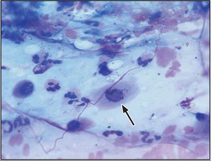
Figure 17.17 Corneal swab from a dog. Scattered neutrophils are observed in a background of mucus. A nonkeratinized squamous epithelial cell coated in melanin granules is indicated by an arrow (Wright–Giemsa, 1,000? magnification).
Inflammation
Inflammation of the cornea can occur secondary to physical or chemical damage, infectious organisms, immune-mediated corneal disease, uveitis, and elevated ocular pressures (Wilcock, 2007). Bacterial and fungal keratitis often causes a suppurative inflammatory response. Fungal keratitis is commonly seen in horses (Cutler, 2004) but has also been reported in dogs and cats (Labelle et al., 2009; Binder et al., 2011; Pucket et al., 2012; Nevile et al., 2016).
Fungal organisms are readily identified in corneal swabs or scrapes. The morphology depends on the type of fungus, but some general features include septation and branching and uniform parallel walls (Figure 17.18). Cytologic specimens from bacterial keratitis are typically highly cellular. Many neutrophils are observed and the neutrophils are often degenerate. Intracytoplasmic bacterial organisms are also seen (Figure 17.19). Recognition of the organisms within the cytoplasm of neutrophils is important to truly recognize them as pathogens rather than contaminants. Within the background are thick strands of mucinous material, which should be differentiated from fungal hyphae. Common bacterial pathogens reported in canine bacterial keratitis include Streptococcus and Pseudomonas spp. (Hindley et al., 2016). The most frequent bacteria isolated from feline bacterial keratitis include the Staphylococcus genus (Golreich et al., 2020). 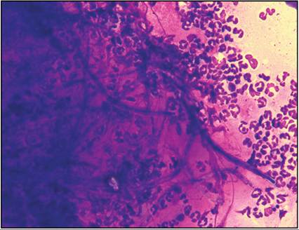
Figure 17.18 Corneal swab from an ulcerative lesion from a feline cornea with fungal keratitis. Neutrophils are observed surrounding fungal hyphae. Septation and branching are noted. Culture revealed Aspergillus flavus (Wright–Giemsa, 1,000? magnification).
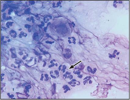
Figure 17.19 Corneal swab from an ulcerative lesion in a dog with septic suppurative keratitis. The sample is cellular, many neutrophils are observed, rarely containing phagocytized rod-shaped bacteria (arrow). Extracellular bacteria are also observed (Wright–Giemsa, 1,000? magnification).
Eosinophilic keratitis has been reported in cats. Feline herpesvirus-1 has been suggested as but not confirmed as a possible etiology (Naisse et al., 1998). Cytologically, eosinophils are seen in high numbers with a mixture of mature squamous epithelial cells, mucinous material, and low numbers of neutrophils (Figures 17.20a, b).
Granules from eosinophils are commonly seen in the background. Mast cells, small lymphocytes, and plasma cells have also been reported in low numbers (Prasse & Winston, 1996; Lucyshyn et al., 2021).Pannus (also called chronic superficial keratitis) is an immune-mediated disease diagnosed most commonly in German Shepherd Dogs and sighthounds. The distinctive clinical appearance in affected breeds includes corneal pigmentation, vascularization, and crystalline degeneration. Because the clinical appearance is highly suggestive of a diagnosis of pannus, cytology is rarely performed in these cases. When cytologic specimens are obtained from the affected areas of the cornea, an inflammatory population consisting of lymphocytes, plasma cells, and few macrophages can be observed.
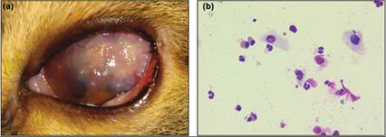
Figures 17.20a,b (a) Domestic shorthair cat with a severe corneal lesion, left eye. (Courtesy Dr. Amber LaBelle.) (b) Corneal swab from the same cat. Fairly equal numbers of eosinophils and neutrophils are observed. A few eosinophils are ruptured, resulting in free granules in the background (Wright–Giemsa, 1,000? magnification).
Neoplasia
Primary neoplasia of the cornea is fairly uncommon; however, SCC, hemangiosarcoma, and melanoma have been reported in multiple species (Kaps et al., 2005; Cazalot et al., 2011; Takiyama et al., 2010; Dreyfus et al., 2011). These tumors exfoliate well and consist of clusters of neoplastic epithelial cells, similar to those diagnosed in the conjunctiva (Figure 17.14). Hemangiosarcoma has been rarely reported (Cazalot et al., 2011). Cytologically, these tumors do not exfoliate well from any tissue, including the cornea, making biopsy a more useful diagnostic tool in these cases. Hemangiosarcoma cells are often spindle-shaped with large round to oval nuclei with prominent nucleoli. Epithelioid variants have been reported with cells described as cohesive (Warren Summers, 2007). Additionally, erythrophagia has been described as a cytologic feature of these tumors (Barger et al., 2012).
Epithelial inclusion cysts have been reported and can be congenital or secondary to previous ocular injury (Simonazzi et al., 2009). Clinically, they appear as white to yellow to tan smooth masses on the corneal surface associated with variable corneal vascularization. Aspiration of these structures reveals aggregates of mature keratinized epithelial cells, primarily anucleate with aggregates of keratinaceous debris. Cytologically, these greatly resemble follicular cysts of the dermis (see Chapter 4).