Intraocular structures
Aspiration of intraocular structures may be deliberate or unintentional. Aspirates of aqueous and vitreous humors are useful diagnostic tools.
Aqueous humor
Aqueous humor is a clear and colorless fluid that has been described as acellular and of low protein concentration in multiple species (Hazel et al., 1985).
Anterior uveitis results in breakdown of the blood–aqueous barrier and transudation of inflammatory cells into the aqueous humor, increasing the cellularity and protein concentration. The ciliary body and iris contain melanocytes, so during an inflammatory process melanin granules and melanophages may exfoliate into the aqueous humor (Raskin, 2010). Causes of uveitis are numerous, including neoplasia, metabolic derangements (diabetes mellitus, systemic hypertension), immune-mediated, infectious (bacterial, protozoal, fungal, parasitic), viral (canine distemper virus, feline infectious peritonitis), traumatic, and idiopathic. Cytologic evaluation of aqueous fluid can aid in the diagnosis of inflammatory uveitis and lymphoma (Wiggans et al., 2013; Wegg et al., 2020). Additional diagnostic tests, such as immunocytochemical staining for feline coronavirus antigen or PCR testing for viral or infectious disease, can be beneficial in obtaining a definite diagnosis (Felten et al., 2018; Wiggans et al., 2013). Neoplasms of the anterior uvea (iris and ciliary body) can be aspirated; however, this type of diagnostic sampling is infrequently performed. Melanoma is a common anterior uveal tumor (Grahn et al., 2006) and consists of individualized and aggregates of highly pigmented neoplastic melanocytes. Ciliary body tumors are most frequently ciliary body adenoma and adenocarcinoma (Wilcock, 2007). These cells appear distinct from melanoma because they exfoliate in tightly cohesive clusters and are not pigmented. The adenocarcinomas will exhibit obvious cellular criteria of malignancy including large prominent nucleoli (Hendrix Donnell, 2007; Ferreria et al., 2019).Vitreous humor
Vitreous humor is a gelatinous fluid of low cellularity, which often contains melanin granules. Inflammation can result in a decreased viscosity of the fluid. Posterior uveitis caused by organisms such as Blastomyces dermatitidis causes severe inflammation, primarily neutrophilic. The fluid is highly cellular and organisms are readily identified (Figure 17.21a). Protothecosis (Figure 17.21b), histoplasmosis, and cryptococcocus can also result in severe uveitis (Schultze et al., 1998; Wilcock, 2007; Raskin, 2010). When aspirating vitreous, other structures in the globe can be aspirated, especially in patients with intraocular diseases such as uveitis, rupture of the lens capsule, or retinal detachment.
id=fig17.21.jpg class="lazyload" data-src="/files/uch_group75/uch_pgroup311/uch_uch7426/image/image989.jpg" alt=fig17.21.jpg>
Figures 17.21a,b (a) Vitreous aspirate from a dog with uveitis of both eyes. A fungal yeast organism (arrow) consistent with Blastomyces dermatitidis is pictured. The background consists of thick stippled proteinaceous material, consistent with vitreous. (b) Vitreous aspirate from a dog with retinal prothecosis. Organisms are noted with few neutrophils (Wright–Giemsa: both, 1,000? magnification). (Courtesy Dr. Reema Patel.)
Lens
The lens consists of an elastic capsule, tightly adherent squamous to cuboidal epithelial cells, and lens fibers (Samuelson, 2007). The lens fibers are elongate structures with scalloped edges that resemble lasagna noodles (Figure 17.22). Aspiration of the lens results in the aspiration of many of these fibers. The retina, especially when detached, can be easily aspirated.
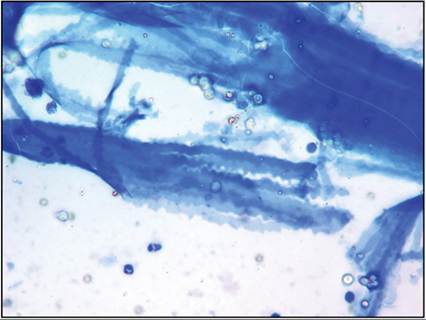
Figure 17.22 Vitreous aspirate from a dog with a luxated lens. Many lens fibers with scalloped edges are present (Wright–Giemsa, 500? magnification).
Retina
The retina consists of ten layers: retinal pigmented epithelial layer, photoreceptor layer, external limiting membrane, outer nuclear layer, outer plexiform layer, inner nuclear layer, inner plexiform layer, ganglion cell layer, nerve fiber layer, and internal limiting membrane.
Aspiration of the retina often reveals a disorganized arrangement of cells. Distinguishing features of retinal cells include the unique shape of the photoreceptor nuclei (Figure 17.23) and the rod-shaped melanin granules of the retinal pigmented epithelium (Figure 17.24). Occasionally, these granules are phagocytized by macrophages or observed in the background (Figure 17.25). Recognition of these structures is very important so that appropriate interpretation of these findings can be made. 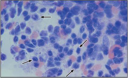
Figure 17.23 Vitreous aspirate from a dog with uveitis caused by Blastomyces dermatitidis. The patient had a detached retina of the right eye. Aspiration of the retina was performed and characteristic photoreceptor nuclei are observed (arrows) (Wright–Giemsa, 1,000? magnification).
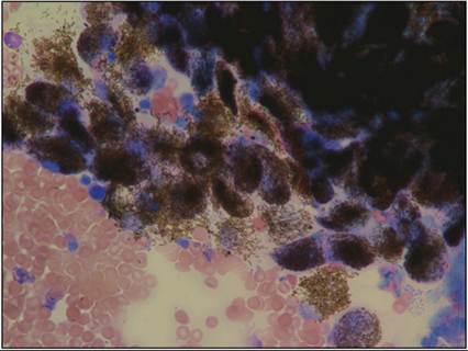
Figure 17.24 Retinal pigmented epithelium from the same sample as in Figure 17.19 (Wright–Giemsa, 500? magnification).
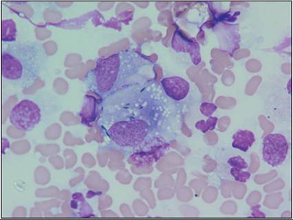
Figure 17.25 Vitreous aspirate from a dog with uveitis. A macrophage, containing many pigment granules likely from the retinal pigmented epithelium, is observed (Wright–Giemsa, 1,000? magnification).