Endocrine pancreas
Normal endocrine pancreas
Endocrine cells of the pancreas are found in small islets that contain multiple types of cells. The most common cell in these islets is the β cell responsible for the production of insulin.
Other cells found in the islets include cells responsible for the production of glucagon, δ cells, which produce somatostatin, and F-cells, which secrete pancreatic polypeptide. Normal endocrine pancreas may be sampled for cytology when collecting fine-needle aspirates or impression smears of the exocrine pancreas. These samples would likely contain low to very low numbers of endocrine pancreatic cells. Endocrine pancreas would be identified as groups of cell-free nuclei on a background of lightly basophilic cytoplasm. The nuclei would be round, exhibit minimal anisocytosis, and have finely stippled chromatin and a single prominent nucleolus. Intact cells should have a high N:C ratio and similar nuclear findings.Endocrine pancreas inflammation
Cytologic inflammation of the endocrine pancreas has not been described. Histologically, inflammation can be observed in the endocrine pancreas secondary to exocrine pancreatitis or immune-mediated isletitis. Immune-mediated disease would be characterized by an infiltrate of lymphocytes and plasma cells (Miller, 2017). A study characterizing pancreatic islet findings noted lymphocytes more frequently in cats with diabetes mellitus compared to controls ( Zi et al., 2016).
Endocrine pancreas neoplasia
Tumors of the endocrine pancreas are most likely to originate from the β islet cells, the most numerous endocrine cells in pancreatic islets (Hawkins et al., 1987), responsible for the production of insulin causing an insulinoma. These lesions are most common in older dogs, average age 9 years. They are found with equal frequency in both limbs of the pancreas and carcinomas are more common than adenomas (Hawkins et al., 1987).
Frequency in the cat is extremely rare (Jackson et al., 2009; Gifford et al., 2020). Diagnosis is often made based on clinical signs, serum biochemistry results, and abdominal imaging. Proliferation of the endocrine pancreatic cells often leads to increased insulin production, which causes clinical signs associated with hypoglycemia (weakness/lethargy, generalized muscle twitching, ataxia, mental confusion, seizures). Serum biochemistry results often include hypoglycemia and hyperinsulinemia (Hawkins et al., 1987) and a presumptive diagnosis is made based on the finding of hypoglycemia during observation of clinical signs and resolution with dextrose therapy. Observation of hyperinsulinemia at the same time as hypoglycemia is also used to make a presumptive diagnosis. Abdominal radiography is often unrewarding due to small tumor size. Abdominal ultrasound may be more beneficial but still not very sensitive, with only 36% tumor identification (Robben et al., 2005). The use of contrast enhanced ultrasonography may be helpful to confirm lesions within the pancreas (Cervone et al., 2019; Burti et al., 2022); however, computed tomography with contrast enhancement has the best sensitivity, 71–96%, to detect primary insulinoma lesions (Buishand, 2022). Adenomas are often found as single lesions 3 cm in size, but cases of multiple adenomas have been reported (La Perle, 2011). Carcinomas are often larger than adenomas, may be multilobular in appearance, and invade the surrounding parenchyma (Miller, 2017).Cytologic findings include a highly cellular sample composed of sheets of nuclei on a lightly basophilic background of cytoplasm and occasional distinct, rounded epithelial cells (Figures 16.7a–c). Intact cells have a moderate N:C ratio, a moderate amount of lightly basophilic cytoplasm with occasional punctate cytoplasmic vacuoles, and a round, often eccentrically placed nucleus. The punctate vacuoles are also noted in the background cytoplasm. The nuclei have finely stippled chromatin and a single, prominent nucleolus.
A higher degree of cellular atypia is expected in a β-cell carcinoma as compared with a β-cell adenoma, but the presence of marked atypia and mitotic figures is rare (Miller, 2017). 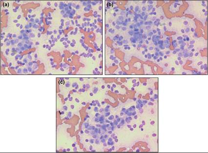
Figures 16.7a–c Insulinoma. Aspirates of an insulinoma confirmed on histopathology. Note the classic bare nuclei appearance and minimal anisocytosis (Wright–Giemsa, 500? magnification).
Common sites for metastasis include the liver and regional lymph nodes (Hawkins et al., 1987) and metastasis can be present in up to 50% of patients at the time of diagnosis (Lurye Behrend, 2001). The presence of metastatic disease makes resolution difficult even though surgical excision of the primary lesion is still recommended. Removal of the primary tumor and debulking of metastatic disease may be enough to cause resolution or lessening of clinical signs (Lurye Behrend, 2001). Prognosis is highly dependent on the presence or absence of metastatic lesions at the time of diagnosis for dogs treated surgically (Lurye Behrend, 2001). Lack of hypoglycemia has been documented in 50% of dogs with no evidence of metastatic disease for up to 14 months after surgery. During the same time frame, lack of clinical signs was documented in 20% of dogs with metastatic disease (Caywood et al., 1988). A median survival of 74 days is reported in dogs undergoing medical management (Tobin et al., 1999).
Immunocytochemical staining with insulin will help to confirm the diagnosis of insulinoma; however, the intensity of staining does not correlate with tumor hormone production, degree of hypoglycemia, or degree of hyperinsulinemia (Hawkins et al., 1987). Endocrine neoplasia of the other pancreatic islet cells has been reported in small animals, but the reports are sparse and cytologic findings are not confirmed.