Chemoreceptor organs
Chemoreceptor tissue is found in multiple locations throughout the body, but tissue in the carotid body and aortic bodies is the most likely to form a mass lesion that may be sampled by cytology.
These tissues are responsible for detecting changes in blood carbon dioxide content, pH, and oxygen tension, and changing respiration and blood flow accordingly.Chemoreceptor organ neoplasia
Canine breeds overrepresented in case series reports for chemoreceptor organ neoplasia include the English Bulldog, Boston Terrier, and Boxer (Hayes & Sass, 1988). Aortic body tumors occur more frequently in dogs, which is in contrast to humans where carotid body tumors are more frequent (Hayes & Sass, 1988; Brown et al., 2003). In a study of 111 cardiac tumors, 31 aortic body tumors (27.9%) were identified (Janus et al., 2016). Aortic body (Buergelt Das, 1968) and carotid body (Yates et al., 1980) tumors have also been described in the cat.
Aortic body tumors appear as a single mass or multilobulated lesion at the base of the heart (~90%) and are less likely to be of mediastinal or sternal/hilar lymph node origin (~10%) (Brown et al., 2003). They may cause clinical signs associated with cardiac compromise and, less likely, respiratory signs associated with deviation/compression of the trachea. In dogs, aortic body adenomas are more common than carcinomas, and criteria for differentiation include tumor size and infiltrative behavior.
Carotid body tumors occur at or near the bifurcation of the common carotid artery, leading to a mass in the cranial cervical or retropharyngeal region. These lesions usually occur in older dogs, with a median age of 10 years (Obradovich et al., 1992). As with aortic body tumors, the distinction between an adenoma and a carcinoma is based on tumor size and local infiltration. Metastasis is common in carotid body carcinomas (up to one-third of cases), and occurs most frequently to the lungs, kidneys, liver, pancreas, and tracheobronchial and mediastinal lymph nodes (La Perle, 2011). A case of metastasis to the submucosal area near the pharynx has also been described in the dog (Hardcastle et al., 2013).
Cytologically, chemodectomas often appear as large numbers of nuclei on a background of basophilic cytoplasm with rare intact cells seen (Figures 16.8a–d). The cells appear to have a small to moderate amount of lightly basophilic cytoplasm with rare punctate cytoplasmic vacuoles and a round, variably placed nucleus. The nuclei have finely stippled to ropey chromatin and one to three small nucleoli. Anisocytosis and anisokaryosis are often mild or, rarely, moderate (Hardcastle et al., 2013). Low numbers of spindle-shaped mesenchymal cells may also be seen. This population exhibits minimal anisocytosis and anisokaryosis. The cells have a small amount of basophilic cytoplasm and an elongate, central nucleus. The nuclei have finely stippled chromatin and inconspicuous nucleoli. Rarely, a small to abundant amount of extracellular, pink, fibrillar matrix may be identified, which may lead to a misdiagnosis of ectopic thyroid tissue or thyroid neoplasia (see Figure 16.5b). Immunochemical stains that may be helpful to confirm the diagnosis of an aortic or carotid body tumor in the dog and cat include neuron-specific enolase, synaptophysin, S-100, and chromogranin A (Brown et al., 2003; Paltrinieri et al., 2004; Aresu et al., 2006). Neuron-specific enolase and synaptophysin staining was not found to correlate with histologic grade, but chromogranin A and S-100 staining intensity and density were found to decrease with increasing tumor grade (Brown et al., 2003; Aresu et al., 2006).
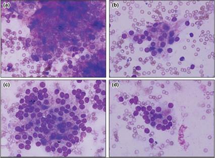
Figures 16.8a–d Chemodectoma. Dog. Aspirates of a mass in the right auricle and right side aortic arch. Histopathology confirmed chemodectoma (Wright–Giemsa, 1,000? magnification).