Adrenal gland
Normal adrenal gland
The adrenal gland is divided into two distinct regions, which appear cytologically different. The adrenal cortex is comprised of the zona glomerulosa, zona fasciculata, and zona reticularis.
Cells of this layer are derived from coelomic epithelium during development, which may be the reason for increased numbers of intact cells on cytology compared with other endocrine organs (La Perle, 2011). Distinction between the layers is not possible on cytology. Normal adrenal cortex cells will appear as large numbers of cell-free nuclei on a background of lightly basophilic cytoplasm with many distinct lipid vacuoles (Figures 16.9a–c). Intact cells have a moderate to low N:C ratio. The nuclei are round, variably placed, and have coarse chromatin and a single, small nucleolus. 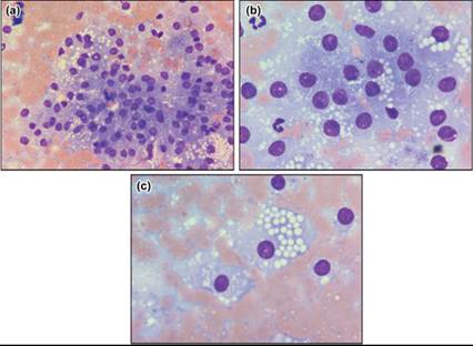
Figures 16.9a–c Normal adrenal cortex. Impression smears of normal adrenal cortex. Note the distinct cytoplasmic vacuoles in many cells, consistent with lipid (Wright–Giemsa: a, 500? magnification; b and c, 1,000? magnification).
Adrenal medullary cells have a classic endocrine appearance as they are derived from ectoderm of the neural crest (La Perle, 2011). Normal adrenal medulla will be highly cellular and contain cell-free nuclei on a background of basophilic cytoplasm. Intact cells have a high to moderate N:C ratio and a round, central nucleus. The nuclei have finely stippled chromatin and inconspicuous nucleoli.
Adrenal gland inflammation
Inflammation within either portion of the adrenal gland has not been previously described cytologically. Pathologically, the adrenal cortex is a common place for inflammation during systemic infections (Miller, 2017). Adrenal gland abscessation has been reported in the dog (Vuong & Aoki, 2021). Lymphoplasmacytic inflammation has been documented in the adrenal glands of dogs and cats with hypoadrenocorticism, with the inflammatory response thought to represent autoimmune disease (Friedenberg et al., 2018; Roberts & Dobromylskyj, 2022).