Enteropathogenic Escherichia coli
Most E. coli isolates recovered from diarrheic rabbits are EPEC strains, which characteristically are found attached to the surface of enterocytes. They produce minimal or no Shiga toxin and are not considered to be enteroinva- sive.
There are significant variations in the pathogenicity of EPEC isolates. Strains of low virulence cause problems primarily in rabbitries with poor sanitation and are usually responsive to antibiotic treatment and improved hygiene. On the other hand, highly virulent strains are often refractory to antibiotic therapy. A large number of strains have been isolated from suckling and weanling rabbits with diarrhea, serotyped, and characterized. In general, these strains of E. coli isolated from naturally occurring diarrheas in suckling or weanling animals produce disease experimentally only in the same age group. For example, strain RDEC-1, isolated from weanling rabbits, attaches only to the enterocytes of weanling rabbits and produces disease in this age group but not in sucklings. This may be due to the absence of the sucroseisomaltose enzyme complex on the enterocyte brush border of suckling rabbits. The enzyme complex develops after weaning and has been shown to permit binding of this strain to enterocytes. In studies of the virulence of strains of E. coli isolated from suckling rabbits, the organism is attached to the enterocytes in both the large and small intestine. In weaned rabbits with experimental coliform enteritis, bacterial attachment occurred in the ileum, cecum, and colon. In rabbits, the adhesins responsible for the attachment of the EPEC strains to enterocytes are an antigenically diverse group.Pathology
The carcass may be dehydrated, and the perineal region is frequently stained with watery yellow to brown fecal material. The cecum and colon are often distended with watery yellow to gray-brown contents.
There may be serosal ecchymoses, edema of the walls of the cecum and colon, edematous mesenteric lymph nodes, and prominent lymphoid tissue in the Peyer's patches and saccu- lus rotundus. Depending on the strain of E. coli, fluid contents may also be present in the small intestine (Fig. 6.34). Microscopically, large numbers of coccoba- cilli are attached to enterocytes in both the small and large intestine. Microscopic changes are normally more extensive in weanlings with the disease. In the small intestine, ileal villi are often blunted, and the lamina propria of affected intestine is edematous, with leukocytic infiltration. Enterocytes at the tips of villi are swollen, and bacteria may be attached to these cells, with effacement of the microvillus brush border. In the cecum and colon, there is bacterial attachment (Fig. 6.35), with variable swelling of affected entero- cytes, and frequently with detachment and erosion involving the tips of the cecal folds. Rabbit EPEC isolates appear to have a predilection for enterocytes overlying Peyer's patches.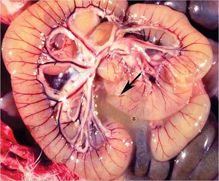
FIG. 6.34. Intestine from a weanling rabbit with profuse diarrhea associated with acute infection with an attaching and effacing enteropathogenic Escherichia coli (EPEC). Note the fluid content that has leaked from an incision in the small intestine (arrow).
Diagnosis
The age, history, clinical signs, and gross and microscopic findings are useful criteria in making the diagnosis. The presence of EPEC is often found as a copathogen in cases of enterotoxemia and other enteritides of the rabbit. The characterization of the isolate of E. coli is recommended in order to determine whether the strain is likely to be a primary pathogen. Isolates of E. coli have been divided into biotypes according to their carbohydrate fermentation patterns. There is a good correlation between biotype and serotype in identified pathogenic strains. Differential diagnoses include clostridial enterotoxemia, Tyzzer's disease, viral enteritides, and acute coccidiosis. Concurrent infection of E. coli and Lawsonia intracellularis has been associated with proliferative enterocolitis.
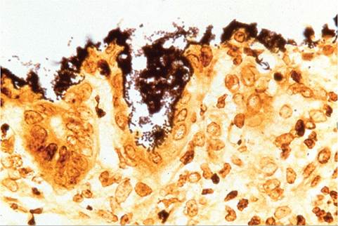
FIG. 6.35. Cecum from a case of acute coliform enteritis in a weanling rabbit. The surface mucosa is densely populated with attaching Escherichia coli (Warthin-Starry stain).