Evaluation of gallbladder fluid
Cholecystocentesis (aspiration of gallbladder content) can be performed to obtain samples for culture and cytology. Samples can be collected laparoscopically, but ultrasound guided percutaneous sampling from right-sided transhepatic or right-ventral abdominal approaches have been reported and can be performed with sedation or light anesthesia (Peters et al., 2016; Webb, 2020).
Retrospective review suggests infrequent self-limiting gallbladder leakage or pericystic fluid accumulation as the most common complications and that these typically have minimal distinct clinical effect, but there are infrequent reports of more significant issues such as gallbladder rupture (Peters et al., 2016; Webb, 2020). It is likely that a perceived increased risk of complication prevented sampling in the more severely affected cases during the retrospective period.Grossly, bile fluid is opaque green-brown and variably viscous or mucoid. While bile fluid should not clot, collection into EDTA has been reported and may be appropriate if there is concern for hemorrhage. Bile fluid is harsh on cellular components; rapid preparation of slides is appropriate. Cytologic preparations can be Romanowski stained direct smears, but concentrated or cytocentrifuged preparations make evaluation of cellularity easier.
Romanowsky-stained cytologic samples of bile fluid typically have a green, blue, or gray background with variable amounts of pale basophilic mucoid material. Crystals can be found, including formed bile crystals and amorphous crystalline material (Figure 9.53).

Figure 9.53 Composite of non-cellular material encountered in bile fluid from dogs. Various forms of granular and crystal material can be found in bile fluid, including blue-black granular material (upper left), brown-green globular crystals (upper right), and orange lanceolate bile crystals (lower left), amorphous granular material (lower right) and nonstaining hemp-seed shaped crystals (center).
Also note the pale basophilic mucoid material (Wright–Giemsa, 1,000? magnification).
Most often, the samples are essentially acellular. When cells are found, they often appear poorly preserved and raise concern that the cytologic preparation may not fully represent the in-vivo environment (Figure 9.54). Collection-associated peripheral blood elements can be present. Noninflammatory cellularity is most often cuboidal to columnar biliary epithelial cells and mesothelial cells but spindled mesenchymal cells and concurrently aspirated hepatocytes can be present (Figure 9.55). In a study of gallbladder cystocentesis samples from 140 dogs and cats, 70% of the cases lacked atypical findings. These often receive the ontologically redundant diagnosis of bile.
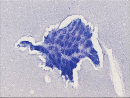
Figure 9.54 Gallbladder aspirate from a 16-year-old castrated male domestic medium haired cat. A sheet of poorly preserved cells is present which may have been biliary epithelium.
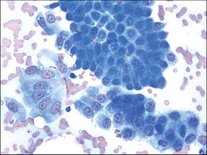
Figure 9.55 Gallbladder aspirate from a 14-year-old castrated male Siamese cat. The sample had more red blood cells and peripheral blood elements than typically encountered in bile fluid. contained sheets of fairly unremarkable cuboidal epithelial cells and fewer columnar epithelial cells (Wright–Giemsa, 1,000? magnification).
When concurrent inflammation and intracellular bacteria are present, a pathologic process is easily diagnosed (Figure 9.56). When one is found without the other, interpretation is more challenging. Suppurative inflammation appears to be the most common type of inflammation but lymphoplasmacytic, eosinophilic, and other forms of inflammation can also be found (Verwey et al., 2021; Peters et al., 2016). Inflammation can be caused by bacteria, protozoa (Isospora spp., Toxoplasma spp., etc., Figure 9.57), liver flukes (Platynosomum spp.), cellular damage, and neoplasia (Palić et al., 2017; Koster et al., 2016).
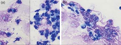
Figures 9.56a,b Gallbladder aspirate from a 16-year-old domestic long-haired cat. (a) Many of the cells in the sample are in poor condition, but neutrophils and rod-shaped bacteria are present. (b) Rarely, phagocytized bacteria are found, helping to diagnose a septic suppurative inflammation.
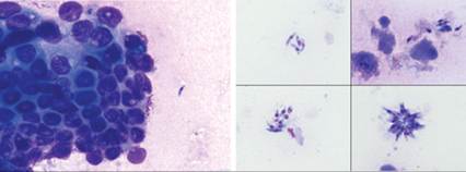
Figure 9.57 Bile fluid aspirate from an immunosuppressed 8-year-old male intact Cocker Spaniel. Extracellular 4–5 um in length apicomplexan zoites are found. These were identified by PCR as Hammondia spp. (modified Wright, 1,000? magnification, images courtesy of Dr. Nora Springer).
The presence of bacteria in bile fluid is called bactibilia. Rod, cocci, and filamentous rod forms can be found on cytology (Figure 9.58). Detection of bactibilia by cytology and culture is not always correlated, and samples with both cytologic and culture confirmed bactibilia will not always have cytologic or histologic evidence of inflammation. One study found that biochemical evidence of hepatic disease or histologically confirmed cholecystitis was diagnosed at a similar frequency in apparently healthy dogs with and without bactibilia diagnosed from a bile fluid sample. This may be because enteric bacteria can transiently be present in bile fluid; advanced detection of bactibilia from healthy dogs commonly found enteric bacteria but a consistent bacterial isolate was not present between patients (Gookin et al., 2023). This same study more commonly identified bacteria in the gallbladder mucus from dogs with a mucocele than healthy dogs but could not distinguish if the bacteria contributed to the mucocele or was a sequalae of biliary obstruction. Although the clinical significance of bactibilia without a clear inflammatory process is uncertain, some authors have argued that bactibilia is an indication for antimicrobial therapy (Palić et al., 2017).
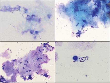
Figure 9.58 The presence of bacteria in bile fluid is called bactibilia. Long rod, cocci, and mixed bacteria can be found. Finding bacteria alone is of unknown significance.
Neoplasia of the biliary tree can be diagnosed by cholecystocentesis, but the diagnostic sensitivity has not been defined in dogs and cats.