Functional anatomy: nose and paranasal sinuses
The nose is the externally visible portion of the respiratory system. It can be divided into an external and internal portion. The external nose consists of the bone, hyaline cartilage, muscle, skin, and mucous membrane protruding from the face.
The immovable portion of the external nose consists of the rostral ends of the nasal bones and the incisive bones. The nasal cartilage extends rostrally from these bones.The external nares (nostrils) are the external openings to the respiratory tract (Fig. 14.1). The philtrum is the area between the lips and nose. It is relatively deep in carnivores and small ruminants, but shallow or absent in the pig, ox, and horse. The pig possesses a rostral bone in the tip of its flattened, cylindricalshaped nose, apparently to assist in rooting. The lateral portion of the nose has sebaceous and sweat glands. The most rostral portion of the nose lacks sebaceous glands, except in the horse.
The nasal cavity extends from the external nares to the caudal nares, and it is separated from the mouth by the hard and soft palates (Fig. 14.2). The hard palate consists of the horizontal portions of the incisive, palatine, and maxillary bones; the soft palate is a musculomucosal extension of the hard palate dividing the rostral part of the pharynx into the oropharynx and nasopharynx.
The space inside the internal nose is the nasal cavity, which is divided into two halves by the median nasal septum. This septum is composed of the vomer, nasal, and ethmoid bones, and cartilage. The nasal cavity is divided into three sections. The vestibule is the most rostral portion located just inside the nostrils. The middle section is filled with the nasal conchae, which are scrolls of bone arising from the lateral wall, and covered with a mucous membrane (Fig. 14.2). Named superior to inferior, the three conchae are dorsal, ethmoidal (middle), and ventral nasal concha. The areas between the conchae are called meatuses, and include the dorsal, middle, and ventral nasal meatus.
The common nasal meatus is located between the median nasal septum and conchae, and it is continuous with the other nasal meatuses.The caudal section of the nasal cavity contains many ethmoturbinates (chonchae of the ethmoid bone). The nasal cavity communicates with the paranasal sinuses,
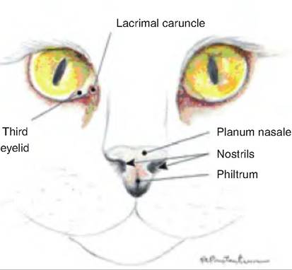
Fig. 14.1. External nose of cat (left) and dog (right). Used with permission from Constantinescu, G.M., 2002. Clinical Anatomy for Small Animal Practitioners. Iowa State Press, Ames, Iowa.
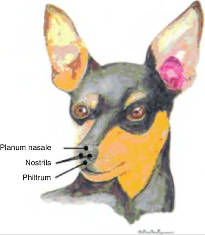
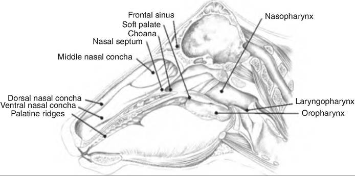
Fig. 14.2. Median aspect of the head of a large ruminant. The dorsal, ventral, and middle nasal concha are visible in the nasal cavity. They are located dorsal to the hard palate and are separated by dorsal, middle, and ventral nasal meatuses. Also shown are the oropharynx, nasopharynx, and Iaryngopharynx. (Reprinted from Constantinescu and Constantinescu, 2004. Used by permission of the publisher.)
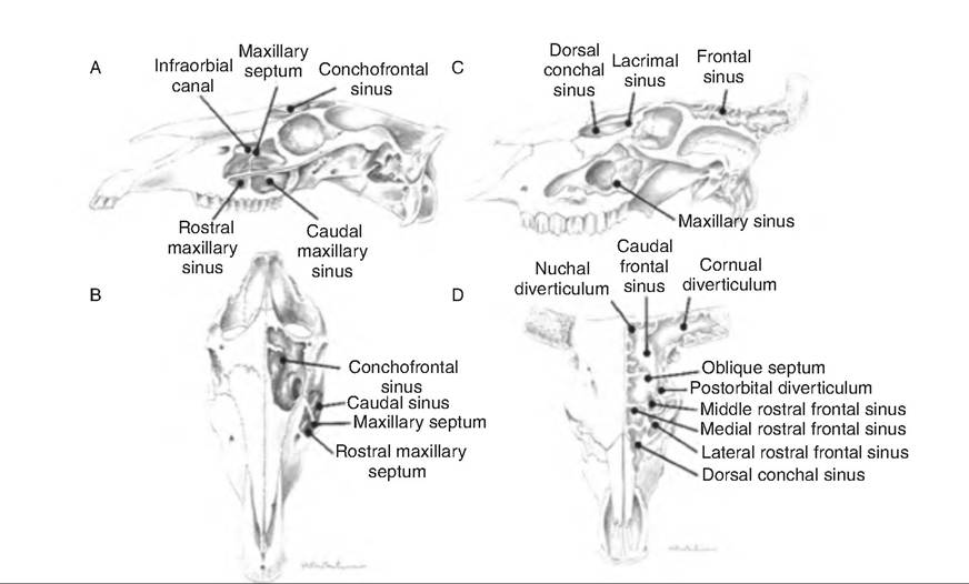
Fig. 14.3. Paranasal sinuses. (A) Lateral aspect of a horse. (B) Frontal aspect of a horse. (C) Lateral aspect of a large ruminant. (D) Frontal aspect of a large ruminant. (Reprinted from Constantinescu and Constantinescu, 2004. Used by permission of the publisher.)
and posteriorly with the nasopharynx through two openings called the internal nares, or choanae (Fig. 14.3). The paranasal sinuses are air-filled cavities within some bones of the skull. The major ones are the frontal and maxillary sinuses, but others may be present depending on the species. In horned cattle, the frontal sinus can extend into the horn as the cornual diverticulum.