Conducting pathway anatomy
Pharynx and nasopharynx
The pharynx connects the nasal cavity and mouth to the larynx and esophagus, respectively. Commonly called the throat, it directs food and air into the digestive and respiratory systems, respectively.
The soft palate divides the rostral portion of the pharynx into the oropharynx and nasopharynx (Fig. 14.2), and the common caudal portion is the Iaryngopharynx.The nasopharynx is located dorsal to the soft palate extending from the internal nares to the Iaryngophar- ynx. The palatopharyngeal arches are found at the border of the nasopharynx and Iaryngopharynx. Since it is located above the mouth, it serves as a passageway only for air. When the animal swallows, the soft palate and uvula move superiorly, closing off the nasopharynx and preventing food and water from entering the nasal cavity.
The pharyngotympanic, or auditory, tubes drain from the middle ear to the nasopharynx. It helps equalize the pressure within the middle ear with atmospheric pressure.
Oropharynx and Iaryngopharynx
The oropharynx lies ventral to the soft palate extending from the oral cavity to the base of the epiglottis. The palatoglossal arches lie at the border between the oral cavity and oropharynx. This is a common pathway for both swallowed food and inhaled air. The Iaryngo- pharynx also serves as a common pathway for food and air. It extends from the epiglottis to the larynx, the diverging point for the respiratory and digestive systems.
Larynx
The larynx connects the Iaryngopharynx with the trachea, and it contains the vocal cords. The two functions of the larynx are (1) to provide a routing mechanism for air and food, and (2) to make sounds. Superiorly, the larynx attaches to the hyoid bone. The larynx is formed by five mucus-covered cartilages including single epiglottic, thyroid, and cricoid cartilages, and paired arytenoid cartilages (Box 14.1).
The epiglottic cartilage provides structure to the epiglottis, which closes the opening to the larynx during swallowing, thus preventing ingested materials from entering the lungs. The thyroid cartilage is the largest cartilage and forms the "Adam's apple" in humans. The cricoid cartilage connects the thyroid cartilage and trachea. The arytenoid cartilages are paired and irregularly shaped. They have a ventral vocal process to which the vocal ligament (vocal cord) is attached. The glottis consists of the vocal ligaments and the slit-like gap between them, the glottic cleft.
Trachea and bronchi
The trachea, or windpipe, is a cylindrical tube extending from the larynx to the bifurcating right and left primary bronchi above the base of the heart. The cervical portion runs from the larynx to the thoracic inlet, while the thoracic portion continues to the bifurcation of the primary bronchi. The thoracic inlet is formed by the first pair of ribs, the first thoracic vertebra, and the cranial parts of the sternum. The trachea consists of four layers: (1) mucosa, which is the deepest layer; (2) submucosa; (3) hyaline cartilage; and (4) adventitia, the most superficial layer composed of areolar connective tissue. The mucosa consists of pseudostratified ciliated columnar epithelium resting on the lamina propria containing elastic and reticular fibers.
The trachea contains a series of dorsally incomplete, C-shaped hyaline cartilage rings. These rings keep the trachea open. The trachea cartilages are united by the annular cartilage, thus making the trachea flexible (Fig. 14.4). The trachealis muscle is smooth muscle connecting the open, dorsal portion of the cartilaginous rings.
The trachea divides into the right and left primary, or principal, bronchi. Like the trachea, the primary bronchi contain incomplete cartilaginous rings and are lined with pseudostratified ciliated columnar epithelium.
Box 14.1 Purring in cats
It is well known that cats purr while content, but they can also purr when injured and in pain.
The mechanism of purring is not well understood. It was once thought that the purr was produced from blood surging through the inferior vena cava, but it is more widely believed that the intrinsic (internal) laryngeal muscles are the likely source for the purr. The laryngeal muscles are responsible for the opening and closing of the glottis (space between the vocal chords), which results in a separation of the vocal chords and thus the purr sound. There is an absence of purring in a cat with laryngeal paralysis. Studies have shown that the movement of the laryngeal muscles is signaled from a unique "neural oscillator" in the cat's brain.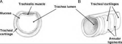
Fig. 14.4. Trachea of a goat. (A) Cross section of trachea. (B)
Annular ligaments. (Reprinted from Constantinescu, 2001. Used by permission of the publisher.)
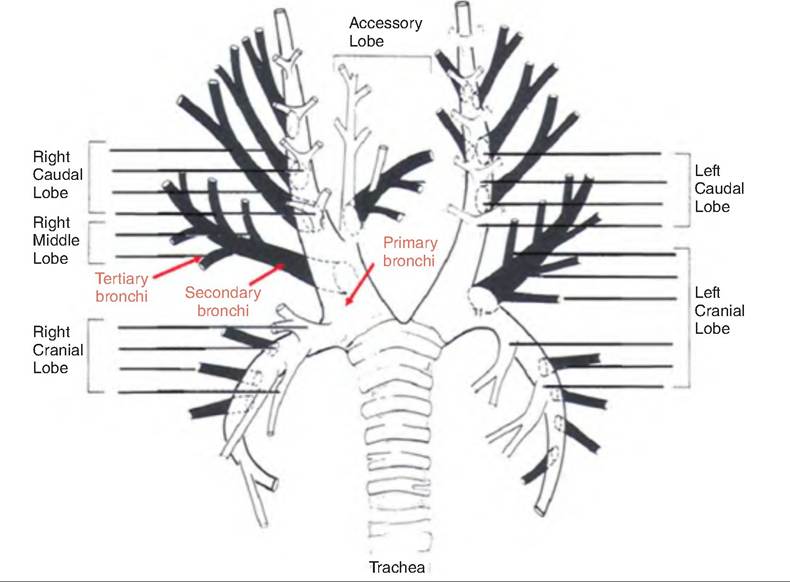
Fig. 14.5. The canine bronchial tree. (Reprinted from Constantinescu, 2002. Used by permission of the publisher.)
Upon entering the lungs at the lung's hilus, the primary bronchi divide into the smaller secondary, or lobar, bronchi. These keep dividing into the following sequence of channels: tertiary, or segmental, bronchi → bronchioles → respiratory bronchioles → alveolar ducts → alveolar sac → alveoli (see Fig. 14.5; see also Fig. 14.7). This extensive branching of the respiratory channels is called the bronchial tree. The alveoli are thin-walled sacs where gas exchange occurs. Below the tertiary bronchi, the mucous membrane changes from ciliated cuboidal epithelium with no goblet cells to nonciliated simple cuboidal epithelium in the respiratory bronchioles.