Respiratory anatomy of the lungs and pleural membrane
The lungs are paired organs located within the thorax. In general, the left and right lungs have two and four lobes, respectively. The horse has three right lobes (Fig. 14.6). The cranial portion, or apex, of each lung is located in the thoracic inlet; the base is the caudal end of the lung resting on the diaphragm.
The hilus of the lung is the medial area where the bronchi, blood vessels, and nerves enter the lungs. The cardiac notch is the indentation between the lobes where the heart makes contact with the lung.The lungs are surrounded by a serous membrane called the pleural membrane. The superficial layer lining the thoracic cavity is the parietal pleura and the layer closely adhering to the lungs is the visceral pleura. The narrow parietal space between these two layers contains a small amount of pleural fluid that allows the two layers to slide over one another during breathing. Inflammation of the pleural membrane is called pleurisy. The mediastinum is the midline site formed where the two pleural membranes meet. It contains the heart, large vessels, esophagus, and other structures, and separates the two lungs from one another.
After the first breath, the lungs become less dense. This fact allows one to determine whether a newborn animal is stillborn. A sample of lung tissue can be placed in water to see whether it floats. If it floats, it indicates that the animal took at least one breath and, therefore, was born alive.
The pressure inside the intrapleural space is negative. This negative pressure is vital for the expansion of the lungs. If an injury to the chest wall punctures the pleural membrane, it can allow air to enter the intrapleural space, resulting in a pneumothorax. Such an injury can be caused by a sharp object penetrating the chest cavity, or a traumatic blow such as being struck by a car. Because this allows the intrapleural pressure to equilibrate with atmospheric pressure, the lung on that side will collapse.
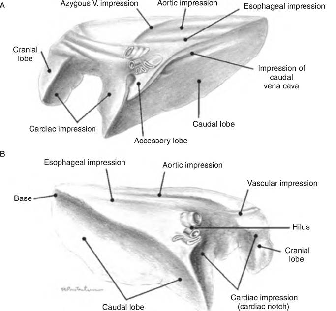
Fig. 14.6. Lungs of a horse. (A) Medial aspect of the left lung. (B) Medial aspect of the right lung. (Reprinted from Constantinescu and Constantinescu, 2004. Used by permission of the publisher.)
Alveoli
Surrounding the alveolar ducts are many alveoli and alveolar sacs. Within the alveolar sac are two or more alveoli, saclike Outpouchings lined with simple squamous epithelium on a thin elastic basement membrane (Fig. 14.7). Alveoli walls contain predominantly type I alveolar cells, which are simple squamous epithelium and are the main site of gas exchange. They also contain type II alveolar (or septal) cells, alveolar macrophages, and fibroblasts that produce reticular and elastic fibers. Type II alveolar cells are cuboidal epithelial cells containing microvilli that secrete alveolar fluid containing surfactant. The alveolar macrophages are wandering phagocytes that remove debris from the lungs.
The respiratory membrane is where O2 and CO2 diffuse across the alveolar and capillary walls. It is a very thin membrane about 0.5 μm thick and consists of four layers:
1. a layer of type I and type II alveolar cells, and alveolar macrophages
2. the epithelial basement membrane
3. the capillary basement membrane
4. the endothelial cells lining the capillary.