18 Gastric adenocarcinoma in a dog
Initial presentation
Vomiting
Signalment: 11-year-old male entire Border collie, body weight 22.1 kg
Case history
The dog had been vomiting intermittently for about 8 months.
For the previous 2 months the frequency of vomiting had increased to at least once a day, usually in the morning. The vomiting occurred either before or after eating. The vomitus usually contained food and bile, but no blood. He was also reported to recently be gulping, retching and drooling. His appetite was still fair, although he was sometimes less interested in food than previously. His faeces were normal in appearance and frequency. He had lost some weight over the past 2 months.He had previously been diagnosed with early congestive heart failure and was on treatment with furosemide (2 mg/kg po q 12 hours) and enalapril (0.45 mg/kg po q 12 hours). His urination and drinking were increased due to the diuretic treatment. His previous signs of weakness, coughing and tachypnoea appeared to be well controlled by the medication.
He was vaccinated yearly, but had not been treated for endoparasites for a couple of years. He was fed three times a day on a diet which included dry dog food as well as fried chicken, pasta, tuna and rice.
Physical examination
The dog was bright, alert and responsive. His body condition score was 4/9. Thoracic auscultation revealed a IV/VI left apex parasystolic heart murmur, with normal lung sounds. Heart rate was 132 beats per minute and he was intermittently panting. His pulse quality was good. The mucous membranes were pink and slightly tacky with a capillary refill time of 200. (The Spec cPL is a quantitative test.) This value is considered to be consistent with pancreatitis. The correlation of the SNAP cPL to the Spec cPL is about 95%.
Urinalysis showed a urine specific gravity of 1.038, consistent with the dog’s ability to concentrate urine when dehydrated, which was also being affected by the use of furosemide (and may have reflected a missed furosemide dose the morning of urine collection).
The urine chemical strip results were unremarkable, with a pH of 7.4, and only a few struvite crystals seen on sediment.Clinical tip
Struvite crystals are normal in dog urine and should not be interpreted as an indication that uroliths (stones) are present. If uroliths are present, then the crystals can be an indicator of the type of stone, but the presence of crystals without stones is not an indication for treatment.
Imaging
A barium impregnated polyethylene spheres (BIPS) study was performed to detect any possible foreign body. A radiograph taken 14 hours after administration showed that all the BIPS had moved out of the stomach into intestines, showing that no obstruction was present (Fig 18.1).
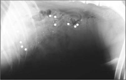
Fig 18.1
Lateral abdominal radiograph showing normal movement of BIPS ruling out a complete gastric obstruction
(courtesy of Dr Tobias Schwarz)
Clinical tip
BIPS are barium impregnated polyethylene spheres packaged with 9 or 10 large (5 mm) BIPS and 30 small (1.5 mm) BIPS. The small ones are meant to mimic the action of small food particles and move through the intestinal tract similarly to food. The large BIPS are similar to non-digestible particles which are only moved into the duodenum during the fasting period from the migrating motility or ‘housekeeper wave’ of contractility. If there is an obstruction present, the BIPS will bunch proximal to it. They do not show gut wall thickness, mucosal detail or outline foreign bodies.
Endoscopy
Gastroduodenoscopy was performed. A large raised mass with a crater ulcer was present at the lesser curvature of the stomach (Fig 18.2). The mucosa around the mass was inflamed, thickened and firm and the mass bled easily when biopsied. The proximal small intestine appeared normal.
Fig 18.2
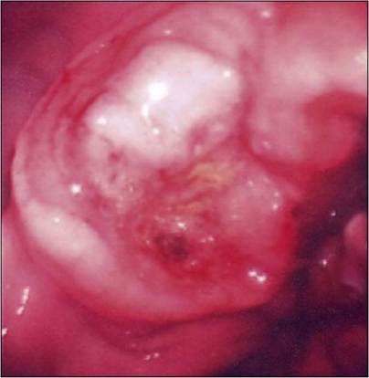
Endoscopic view of raised ulcerated lesion on lesser curvature of the stomach
As neoplasia was suspected, thoracic radiographs were taken.
There was no evidence of pulmonary tumour metastasis present on the radiographs.Histopathology of the samples of the gastric mass was described as gastric ulceration with associated intense granulation reaction. As there was a suspicion of neoplasia, a laparotomy was performed and a surgical biopsy was taken. The histopathology of this biopsy sample was interpreted as gastric adenocarcinoma.
Treatment and outcome
Gastric neoplasms are generally treated by surgical resection; unfortunately in this case the surgeons determined that the extent of the tumour was too great to excise. Chemotherapy for gastric adenocarcinomas is generally unrewarding. In humans with some types of gastric tumours, tumour growth is enhanced by histamine and histamine 2-receptor blockers such as ranitidine or cimetidine may inhibit tumour growth.
Clinical tip on gastrointestinal tumour biopsy
These cancers often cause an inflammatory reaction and biopsies taken only from the mucosa may be interpreted as inflammatory or, if the tumour is affecting the submucosa or muscularis, the mucosa may appear fairly normal. The gastric wall will often feel rigid and stiff and be difficult to distend. If the gastric biopsy results from samples obtained via the endoscope are not consistent with the appearance of the stomach, a full thickness sample obtained by surgery may be required. As the stomach wall is abnormal, this must be done with care and skill, as dehiscence is possible when working with diseased tissue.
Prior to obtaining the second biopsy results this dog was treated with palliative care using ranitidine and sucralfate. As no further treatment was available, the owners elected to euthanize the dog.
Epidemiology
The prevalence of gastric cancer is thought to be low in dogs and cats, accounting for less than 1% of canine tumours. Of the gastric tumours, malignant adenocarcinoma is the most common in dogs and comprises 47 to 72% of gastric malignancies. Many British clinicians believe that the prevalence may be higher in the United Kingdom, although that has not been documented.
While gastric adenocarcinomas or adenomas are the most common gastric tumour in dogs, leiomyomas and lymphosarcoma, as well as leiomyosarcoma, polyps, fibromas, fibrosarcomas, squamous cell carcinomas and plasmacytomas also occur. Some of these tumours have a better outcome than gastric adenocarcinomas, so the tumour type should be characterized. Primary gastric adenocarcinomas are frequently located on the incisura angularis of the lesser curvature (as was this one) or the pyloric antrum of the stomach, although gastric ulcers from other causes may occur here as well. There are three morphological patterns of distribution of gastric carcinomas: diffusely infiltrating non-ulcerating lesions that involve most of the stomach that cause a tough, rigid (leather bottle) appearance, localized, raised thickened plaques usually containing a raised central ulcer (as in this case) and raised, polypoid, sessile lesions projecting into the lumen of the stomach.As with many tumours, primary gastric tumours occur more frequently in older dogs, usually around 8 to 10 years of age with a peak incidence of gastric carcinomas at 11 to 12 years of age, similar to this patient. Male dogs are affected more frequently than female dogs and a higher risk has been suggested for Rough collies, Staffordshire terriers and Belgian shepherd dogs.
About 70% of gastric tumours are malignant and these spread to the regional lymph nodes, liver and lungs. Before surgical treatment is undertaken, the lungs should be checked for evidence of metastasis.
In humans, diet is thought to influence the development of gastric tumours. The prevalence of the tumours in dogs is so low that dietary influences would be difficult to study. Gastric tumours in humans are also related to the presence of Helicobacter spp., but no association with this has been made in dogs.
Prognosis
The prognosis for most patients with malignant gastric cancer is poor. Even in those dogs in which surgery can be performed, most patients are dead within 6 months due to recurrent or metastatic cancer.