19 Pancreatitis in a cat
Initial presentation
Vomiting, poor appetite and lethargy
Signalment: 14-year-old male neutered Burmese cat, body weight 4.4 kg
Case history
The cat had been vomiting for 2 days and was refusing to eat.
The owner had tried to encourage him to eat, but he refused any type of food. He was very lethargic and the owner thought that he had been urinating larger volumes than usual because the litter box was very wet and she had observed this cat in the litter box frequently.The owner had owned the cat since he was a kitten. He lived with a littermate sibling, who was clinically healthy. Both the cats had been vaccinated approximately 18 months earlier, but had not been treated for endoparasites for several years. They were both primarily indoor cats, although occasionally had access to a fenced garden. Neither cat hunted nor scavenged food. They did play with toys, although the patient had arthritis in both elbows, which decreased his mobility. His previous diet was a combination of a commercial dry cat food and a commercial canned cat food.
Physical examination
Physical examination revealed the cat to be quiet and dull, but responsive. He was in slightly overweight body condition (body condition score 6/9). His oral mucous membranes were mildly jaundiced and tacky, with a capillary refill time of 2 seconds. He appeared approximately 7 to 8% dehydrated.
The sclera was mildly jaundiced bilaterally with no other ocular changes noted. Examination of the oral cavity, ears and nose showed no abnormalities. All peripheral lymph nodes were within normal limits. Auscultation of the lung fields as well as compression and percussion of the thorax were normal. The respiratory rate was elevated at 60 breaths per minute. Cardiac auscultation revealed a heart rate of 180 beats per minute with matched pulses. Rectal temperature was 39.1° C.
Abdominal palpation indicated marked cranial abdominal pain. The reminder of the physical examination was unremarkable.Problem list and discussion of problems
The cat’s problems included vomiting, poor appetite and lethargy. The poor appetite and lethargy were thought to be related to the cause of the vomiting. He also was suspected to have polyuria, although it was possible that the other cat in the household had increased urine output. He was also jaundiced.
Differential diagnosis
Differential diagnoses for vomiting
• Dietary sensitivity (allergy or intolerance)
• Dietary indiscretion
• Stomach
• gastritis
• gastric ulcers
• inflammatory bowel disease
• foreign body
• parasites
• neoplasia
• Small intestinal disease
• infectious, e.g. bacterial, viral, fungal
• gastrointestinal parasites
• inflammatory bowel disease
• infiltrative neoplasia, e.g. lymphoma, mastocytosis
• high partial obstruction, e.g. intussusception, foreign body
• Systemic disease
• uraemia
• liver failure
• sepsis
• congestive heart failure
• acidosis
• hypoadrenocorticism
• ketoacidotic diabetes mellitus
• hyperparathyroidism
• gastrinoma
• Neurological diseases (no other signs consistent with these in this cat)
• dysautonomia
• vestibular disease
• CNS disease
• Drugs, toxins (no known history of drug or toxin exposure)
Differential diagnoses for polyuria
• Diabetes mellitus
• Acromegaly (usually concurrent with diabetes mellitus in cats)
• Chronic kidney disease (sometimes acute renal failure) - the signs were too acute for chronic, unless the owner had not observed a gradual increase in urine output
• Pyelonephritis
• Electrolyte imbalances, e.g. hypercalcaemia, hypokalaemia, hyperkalaemia
• Hyperthyroidism
• Liver disorders
• Polycythaemia
• Drugs (no history of or known access to drugs or medications)
Other causes of polyuria/polydipsia, which are uncommon to extremely rare in cats, include:
• Hyperadrenocorticism
• Primary hyperaldosteronism
• Hypoadrenocorticism
• Tubular disorders (e.g.
renal glucosuria)• Central diabetes insipidus
• Psychogenic polydipsia
Differential diagnoses for jaundice
These include pre-hepatic disorder, hepatic disorders and post-hepatic disorders.
• Pre-hepatic jaundice is caused by intra- or extravascular haemolysis
• Hepatic causes of jaundice are due to liver disease, e.g.
• cholangiohepatitis complex
• hepatic lipidosis
• diffuse neoplasia
• feline infectious peritonitis
• toxins, e.g. paracetamol (acetaminophen)
• cirrhosis
• idiopathic drug reactions
• Post-hepatic causes of jaundice are caused by biliary obstruction or rupture,
e.g.
• pancreatitis
• enteritis
• biliary neoplasia
• calculus
• ruptured bile duct or gallbladder
Case work-up
Intravenous fluid therapy with crystalloids at a rate to correct 8% dehydration was initiated.
Minimum data base
Haematology revealed a mild leukocytosis due to mild neutrophilia (13.52 ? 109∕l; reference range: 2.5-12.5 ? 109∕l), a left shift, with 1.5 ? 109∕l band neutrophils and moderate monocytosis (3.07 ? 109/1; reference range: 0.15-1.7 ? 109/l). A mild normocytic, normochromic anaemia was present (PCV: 0.237 1/1; reference range 0.30-0.45 1/1). A blood smear ana1ysis revea1ed the presence of toxic neutrophi1s and reactive 1ymphocytes.
Serum biochemistry revealed mild hypoalbuminaemia (20 g/1; reference range 23-39 g/1) and hyperbilirubinaemia (20 iimol/1; reference range 0-15 iimol/1), elevated alanine aminotransferase (ALT) of 104IU/1 (reference range 6-83IU/1) and moderate hypokalaemia (2.0 mmol/l; reference range 2.9-4.2 mmol/l). Alkaline phosphatase (ALP) was within the reference range at 35 IU/l (reference range 10-100 IU/l). Serum urea and creatinine were both elevated at 17.2 mol/1 (reference range 2.9-9.8 mmol/l) and 161 iimol/1 (reference range 40-177 pmol/l). Hyperglycaemia (26.9 mmol/l; reference range 3.94-8.83 mmol/l) was also present and serum fructosamine was 471 iimol/1 (reference range 159-295 pmol/l).
Cholesterol was mildly increased at 5.5 mmol/l (reference range 2.0-3.4 mmol/l) and triglycerides were increased at 3.3 mmol/l (reference range 0.57-1.14 mmol/l).
Urinalysis revealed a urine specific gravity (USG) of 1.014 and dip stick analysis was 1+ positive for protein, 2+ positive for blood, bilirubin and leukocytes and 3+ positive for glucose. Ketone bodies were not detected. Sediment examination showed the presence of occasional red blood cells, numerous white blood cells and bacteria (cocci) and a sample submitted for bacterial culture was later found to be positive for Staphylococcus.
The cat was diagnosed at this point with diabetes mellitus and a urinary tract infection. The azotaemia present was thought to be due either to pre-renal causes (dehydration) or kidney disease or a combination of both.
Imaging
Abdominal ultrasonography revealed an enlarged hypoechoic pancreas surrounded by a small amount of free abdominal fluid (Fig 19.1). The duodenum appeared corrugated (Fig 19.2), which is often seen with pancreatitis. The liver appeared hyperechoic, but was homogenous and normal in size. There was loss of demarcation between cortex and medulla of both kidneys (Fig 19.3). The right kidney was of similar appearance and contained a small cyst within the cortex. A large amount of sediment was seen in the urinary bladder.
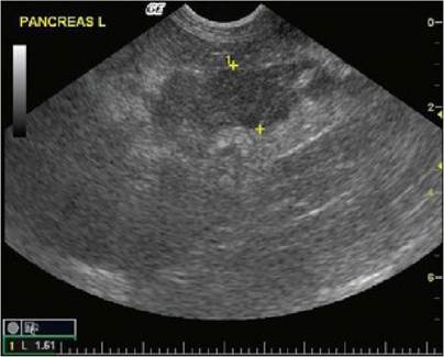
Fig 19.1
Ultrasound picture of pancreas showing uneven appearance
(courtesy of Carolina Urraca del Junco)
Fig 19.2
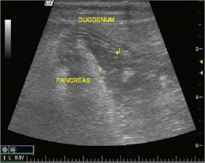
Ultrasound picture of duodenum showing corrugated appearance
due to pancreatitis
(courtesy of Carolina Urraca del Junco)
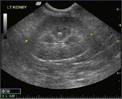
Fig 19.3
Ultrasound of kidney showing decreased corticomedullary junction
(courtesy of Carolina Urraca del Junco)
The enlarged hypoechoic pancreas with free abdominal fluid was considered the clinically most significant finding and was consistent with pancreatitis, with evidence of chronic kidney disease or renal age related changes.
The changes in the liver may have been due to a degree of hepatic lipidosis, which is often seen with diabetes mellitus. The small cyst in the right kidney was thought to be an incidental finding.Further testing
Serum was submitted for a feline pancreatic lipase immunoreactivity (fPLI) test and cobalamin concentrations. The fPLI was increased at 16.9 μg∕l (reference range 0.1-3.5 μg∕l), consistent with a diagnosis of pancreatitis. Serum cobalamin concentration was within the reference range at 946 ng/l (reference range 290-1499 ng/l).
The elevated fPLI plus the ultrasonographic changes in the pancreas confirmed an additional diagnosis of pancreatitis. The jaundice was thought to be post-hepatic in origin and due to the pancreatitis.
Clinical tip on serum cobalamin in cats
Cobalamin is absorbed in the ileal part of the small intestine and requires prior binding with intrinsic factor, which comes from only the pancreas in cats, while in dogs it comes from both the stomach and pancreas. Because of this, cats with pancreatitis are at risk for hypocobalaminia. Other causes of cobalamin deficiency include poor absorption due to intestinal disease, exocrine pancreatic insufficiency, excessive bacteria in the upper small intestine utilizing the vitamin or a deficiency in intrinsic factor.
Treatment and outcome
The cat was stabilized with intravenous fluids (0.9% saline supplemented with potassium chloride at a rate to correct dehydration). Analgesia was provided using buprenorphine (initially at 20 μg∕kg iv q 8 hours, later 10 μg∕kg iv q 8 hours). Maropitant was administered as an antiemetic (1 mg/kg sc q 24 hours) and ranitidine (2 mg/kg sq q 12 hours) was used as an antacid and promotility agent. Clavulanate-potentiated amoxicillin (20 mg/kg iv q 4 hours for 2 days, then 15 mg/kg po q 12 hours) and clindamycin (5.5 mg/kg po q 12 hours) were administered. He also received 1 IU of Caninsulin subcutaneously every 12 hours.
Clinical tip on maropitant use in cats
While maropitant is not approved in cats (at the time of writing), studies have shown that at 1.0 mg/kg it is highly effective in preventing motion-induced emesis in cats. These studies indicate that the NK-1 receptor antagonist maropitant is well tolerated, safe and has excellent anti-emetic properties in cats.
The cat responded very well to this treatment. His demeanour improved and there was no longer evidence of abdominal pain. He accepted syringe feeding very well and no further vomiting was noted.
Repeat measurements of electrolytes were performed and showed improvement and resolution of the hypokalaemia. Blood glucose levels ranged from 10.9 to 18.5 mmol/l.
The cat was discharged on 1 IU of Caninsulin sq q 12 hours, a high protein, low carbohydrate diet and 2 weeks worth of the antibiotics. At a re-visit 1 week later, his blood glucose was 11.2 mmol/l and other haematological and most serum chemistry parameters had improved, although a moderate azotaemia was still present, with a urea of 14 mmol/l and a creatinine of 132 pmol/l. His USG was 1.016, there were no ketones present and the sediment was ‘inactive’. A repeated urine culture was negative.
Nursing tip on examination of urine sediment
To check urine sediment, centrifuge the urine sample for 3 to 5 minutes at 2000 to 3000 rpm. Remove all but a small volume (0.25-0.5 ml) with a pipette or by pouring off the fluid on top. Re-suspend the sediment in the remaining supernatant by gently tapping the tube with your finger. Overzeal- ous shaking may break cells and casts. Transfer a drop of the re-suspended sediment to a microscope slide and cover it with a cover slip. Decrease the microscope light intensity by lowering the condenser and partially closing the diaphragm. Scan the entire sample via the low power objective and then record the average number of elements (e.g. cells, crystals, casts, bacteria) per several high-power fields. Use of stains on wet preparations is by personal preference. Air-dried urine sediment slides may be stained for additional microscopic evaluation.
Urine collected by cystocentesis normally contains only a few cells and sometimes some struvite or oxalate crystals. When there is nothing abnormal in the sediment it is sometimes termed ‘inactive’. Cells seen in healthy animals include zero to three white blood cells and zero to three red blood cells per high powered field and a small number of epithelial cells. A few struvite or oxalate crystals may also be seen in the urine of healthy animals and entire male animals may have some sperm in their urine. Abnormal findings include seeing more white or red blood cells, more than two hyaline casts per high powered field, any bacteria (from a cystocentesis sample) or any other types of crystals.
The cat was monitored every 2 to 3 weeks for the next 6 months. His insulin requirements decreased over time and at the 6 month revisit appointment he no longer required insulin injections. He was relatively healthy without recurrence of pancreatitis or diabetes mellitus (Fig 19.4) until 18 years of age when he was euthanized due to the effects of chronic kidney disease (CKD) and debilitating arthritis.
Fig 19.4
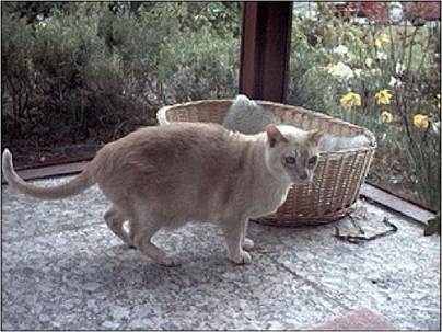
The patient at 16 years of age (2 years after diagnosis)
Nursing tip on nutrition
This cat’s dietary requirements were complicated. High protein diets have been recommended for cats with diabetes, however, as high protein diets are generally high in phosphate and he may have had early chronic kidney disease (CKD), this possibly had to be used with caution. Monitoring his serum phosphorus would be a good indicator for adjusting the diet or addition of a phosphate binder to his diet.
A low fat diet was indicated as he was overweight and extra body weight contributes to insulin resistance and aggravates arthritis. Losing weight is extremely difficult on a high fat diet. With the possibility of early CKD, the use of non-steroidal anti-inflammatory drugs (NAIDs) is also questionable, so he was treated with glucosamine and omega-3 fatty acids. Omega-3 fatty acids have been shown in the dog to decrease the required dose of NSAIDs and also appear to be beneficial in other inflammatory states. Glucosamine has been reported to increase insulin resistance; however, this is usually not a clinical problem as the doses used in the rodent trial showing this were much higher than used clinically.
Discussion and epidemiology
This cat’s combination of urinary tract infection and pancreatitis may have predisposed him to developing diabetes mellitus. He was also overweight, which increases the risk, and a Burmese cat. Burmese cats in the United Kingdom, New Zealand and Australia have a much higher risk of developing diabetes mellitus than other breeds. The development of diabetes mellitus implies that there was possibly chronic inflammation of the pancreas, which had been destroying the function. Pancreatic inflammation is a not an uncommon finding in cats, with significant pancreatic lesions seen in 1.5 to 3.5% of cats at necropsy.
Pancreatitis often occurs in conjunction with diseases of other organs in cats including the liver, kidney, intestine, endocrine pancreas and lungs. In most cases of pancreatitis in cats, an underlying cause or instigating event cannot be determined, leading to classification as idiopathic. Abdominal trauma, infectious diseases (toxoplasmosis, liver flukes, FIP and FIV), lipodystrophy and organophosphate administration have been associated with feline pancreatitis, but appear to account for a very small percentage of cases. Hypercalcaemia precipitated pancreatitis in one experimental study in cats and aspirin induced pancreatic cell damage in another. Potential risk factors for pancreatitis in dogs, such as obesity, dietary indiscretion, a high fat meal, high fat diets and pre-existing endocrine diseases (diabetes mellitus, hyperadrenocorticism, hypothyroidism and hyperlipidaemia) do not appear to be risk factors in cats. It is not known whether dietary factors, e.g. a high fat diet, predispose to pancreatitis in cats.
Inflammatory bowel disease (IBD) may be an important risk factor for the development of pancreatitis (and inflammatory liver disease) in cats, although there was no evidence of that in this cat, other than the vomiting, which is the most common clinical sign of IBD in affected cats. Vomiting can raise intraduodenal pressure and predispose to reflux of enteric contents into the pancreatic duct. Cats may be more susceptible than dogs to pancreatic pathology as a result of such reflux because the feline sphincter of Oddi is a common channel for the pancreatic and biliary outflow at the duodenal papilla and cats have much higher numbers of bacteria in the proximal duodenum than dogs (108 vs. 104 organisms/ ml).
Pancreatitis is classified as acute or chronic (relapsing). Acute pancreatitis is further sub-classified as acute necrotizing or suppurative. Pancreatic biopsy samples for histopathology are required for accurate classification and this cat, like many patients with pancreatitis, was not a good candidate for anaesthesia. Both acute and chronic pancreatitis may be involved in the development of transient or permanent diabetes mel- litus in cats, as in this cat. Some cases of acute pancreatitis are associated with severe clinical syndromes including shock, disseminated intravascular coagulation and multi-organ failure, which he fortunately did not appear to develop.
No significant age or gender predisposition has been recognized in cats with pancreatitis. This varies from the ‘fat, five and female’ concept of pancreatitis in dogs. In cats, a wide age range for pancreatitis (5 weeks to 20 years) has been reported, but some authors suggest that cats older than 7 years are more likely to be affected.
Lethargy (86-100%) and partial to complete anorexia (95-97%) are the most common clinical signs in cats diagnosed with pancreatitis. All other clinical signs occur only sporadically. Classic signs of pancreatitis, such as vomiting and abdominal pain, which occur in dogs are usually absent in cats. Vomiting occurs in about 35% of cats compared to 90% of dogs, abdominal pain occurs in 25 to 52% of cats compared to 58% of dogs, diarrhoea in 15% of cats compared to 33% in dogs and fever in 7% of cats compared to 32% in dogs. Other reported clinical findings in affected cats include dehydration (92%), tachypnoea (74%), hypothermia (68%), tachycardia (48%) and dyspnoea (20%). Almost one-quarter (23%) of cats with pancreatitis have a palpable abdominal mass that may or may not appear painful and can be easily misdiagnosed as a lesion of other intra-abdominal structures such as intestinal tract or mesenteric lymph nodes. Tachypnoea or dyspnoea may occur due to pain, but pleural effusion and pulmonary thrombosis have been diagnosed concurrently with pancreatitis in cats and should not be overlooked as possible causes. In one study of 21 cats with pancreatitis, 66% had complicating conditions.
Diagnosis
The diagnosis of pancreatitis in cats can be challenging. Haematologic abnormalities are uncommon and non-specific and may include a mild anaemia that may be non-regenerative and a leukocytosis, possibly with a left-shift or toxic white cells. Other hematologic changes reflect fluid loss and haemoconcentration. Serum chemistry abnormalities include mild to moderate elevations in ALT, ALP and bilirubin and may reflect concurrent hepatic disease (hepatic lipidosis or cholangiohepatitis) or biliary stasis. Azotemia may be pre-renal, secondary to dehydration, in many cases, although underlying chronic kidney disease should also be considered. Hyperglycaemia is commonly seen in cats due to concurrent stress or diabetes mellitus. Hypocalcaemia is occasionally seen due to saponification of peripancreatic fat.
Changes on abdominal radiographs are often subtle and subjective. Decreased contrast in the anterior abdomen, dilated and gas filled small intestines and transposition of the duodenum, stomach and colon are commonly reported.
Abdominal ultrasound may reveal an enlarged hypoechoic pancreas surrounded by hyperechoic mesentery, with or without dilated pancreatic ducts. Ascites is occasionally observed. The sensitivity of ultrasound is obviously operator and equipment dependent, with reported sensitivities ranging from 35% to 75%. These results underscore the point that a normal ultrasound does not rule out pancreatitis. The clinician should also consider differential diagnoses aside from pancreatitis, including pancreatic neoplasia and pancreatic oedema (associated with hypoproteinaemia or portal hypertension). Computed tomography has not shown any benefit compared to ultrasound for the diagnosis of pancreatitis.
Serum lipase and amylase activities are nearly useless in the diagnosis of pancreatitis in cats, with serum concentrations being neither specific nor sensitive. Determination of serum trypsin-like immunoreactivity (fTLI) measures antibodies against circulating trypsin and trypsinogen. TLI is cleared by the kidneys, so elevations can occur with renal dysfunction. Increases in TLI, which can be quite high, often return to the reference range within 2 to 3 days.
fPLI is more sensitive than any other non-invasive test for the diagnosis of feline pancreatitis and is the test of choice for diagnosis. As it stays elevated while there is ongoing inflammation, it appears to be useful for both acute and chronic pancreatitis.
The theoretical ‘gold standard’ for diagnosing pancreatitis is pancreatic biopsy for histologic evaluation. Peripancreatic fat necrosis is a typical finding in cats with pancreatitis, with variable amounts of pancreatic acinar cell necrosis and inflammation. Chronic pancreatitis is characterized by interstitial fibrosis with acinar atrophy and lymphocyte infiltrates. The disease can have a ‘patchy’ or multifocal distribution and pancreatic biopsies should always be procured during laparotomy even if the gross appearance of the organ is normal. Unfortunately, the lack of pancreatic inflammation on a single biopsy does not rule out pancreatitis conclusively.
Therapy
The old concept of nil per os or withholding food from patients with pancreatitis has little evidence for effectiveness and in humans is thought to possibly have negative consequences for outcome. In cats that may have hepatic lipidosis, nutritional support is especially important. In cats that are not vomiting, feeding a palatable diet is recommended, although anorexia is a common problem. In cats that are not vomiting but still not willing to eat, placement of a nasogastric/naso-oesopha- geal tube is a good alternative. There is no evidence that feeding a low fat diet is beneficial in cats with pancreatitis, so most liquid diets formulated for cats can be used. Anti-emetics are also useful and may also decrease the nausea associated with pancreatitis in cats that are not eating even though they are not vomiting. In cats that are vomiting, ideally a jejunostomy tube would be placed for nutritional support; however, due to the anaesthesia required for this procedure this is often not feasible.
Abdominal pain may be present in cats with pancreatitis even if the cat is not showing obvious signs. Cats often mask signs of pain. Routine analgesia is recommended, as the previous concerns that morphine and related drugs may exacerbate pancreatitis have been discounted.
In dogs, intravenous plasma has been recommended to replete the alpha-macroglobulin, a scavenger protein for activated proteases, which is depleted in pancreatitis. While in humans, the use of plasma has not shown improved survival, many clinicians believe that it is useful. Fresh frozen plasma can be used if available or whole blood (after blood typing the cat and using matched blood) may be used if it is not.
The use of antibiotics in pancreatitis is controversial. There is no evidence that infectious agents cause pancreatitis, however, many clinicians believe in using antibiotics, especially in cats. One rationale is the potential for bacterial infection of the biliary system and pancreatic duct because of the direct communication of the common bile duct and pancreatic duct in the cats. Some clinicians also feel that corticosteroids are useful in cats with chronic (but not necessarily acute) pancreatitis, but this is also controversial, especially in a case with concurrent diabetes mellitus.
Prognosis
The prognosis for cats with pancreatitis depends upon the severity of the disease and the extent of any pancreatic necrosis, the complications and the presence of concurrent disease. Some chronic cases are subclinical and may flare up from time to time. Some patients with chronic disease may develop diabetes mellitus, as did this case, and some have also been reported to develop exocrine pancreatic insufficiency, although the incidence of these sequelae has not been reported. In cats which develop diabetes mellitus, a percentage of them are able to eventually maintain their blood glucose without insulin, a state termed diabetic remission. Some of these will relapse and need insulin, but may be able to achieve another remission with insulin therapy and careful monitoring.