Introduction
As biology students learn early in their schooling, the presence of mammary glands distinguishes mammals from other animals. With a few exceptions, for example, bottle-fed human infants or early-weaned dairy calves, lactation is critical for survival of the neonate and ultimately reproductive success.
Regardless of the specific arrangement or number of mammary glands, milk synthesis and secretion requires development of a functionally mature mammary gland. In reproductively competent mammals, the mature mammary gland consists of a teat or nipple, associated ducts which provide for passage of milk to the outside, and alveoli composed of secretory epithelial cells and supporting tissues. The epithelial cells are arranged to form the internal lining of the spherical alveoli, and these epithelial cells synthesize and secrete milk. Secretions are stored within the internal space of the hollow alveoli and larger ducts between suckling episodes (see Fig. 18.6). Milk is a complex fluid. Consequently, understanding lactation requires multiple disciplines: histology and cytology, biochemistry, endocrinology, cardiovascular physiology, and metabolism to name a few. In addition, the fully functioning mammary gland places striking demands on the physiology of the lactating mother (Tucker, 1994, 2000).Development of the mammary gland during gestation and subsequent differentiation of alveolar cells to allow onset of milk synthesis and secretion in correspondence with parturition is a biological marvel. Mammary secretions first appear as colostrum and subsequently as mature milk and provide the neonate with a spectrum of all the nutrients necessary for good health and early development. Milk of all mammals contains variable amounts of proteins, carbohydrates, and fats suspended in an aqueous medium. Although there are species differences in milk composition, having the birth of the neonate and functionality of the mammary gland coincide is critical.
Among scientists, the study of lactation and mammary development provides a rich resource for cell biologists, endocrinologists, nutritionists, cancer researchers, dairy specialists, and others (Akers, 2002; Rowson et al., 2012). The goal of this chapter is to provide an overview of mammary development and function (Box 18.1).Anatomy and Physiology of Domestic Animals, Second Edition. R. Michael Akers and D. Michael Denbow. © 2013 John Wiley & Sons, Inc. Published 2013 by John Wiley & Sons, Inc.
Given the variety of mammals and the environmental niches occupied, it is no surprise that there is much
Box 18.1 Lactation in males?
There are scattered reports in the scientific literature of mammary development and appearance of mammary secretions in males. In most instances, it is likely most appropriate to characterize this development and appearance of secretion as gynecomastia and galactorrhoea, respectively. In most reported cases, causes have been linked to hormonal imbalances (humans) or animals feeding on plants with estrogenic compounds. However, there are at least two reports from field observations of wild male fruit bats exhibiting overt signs of mammary development and expression of mammary secretions (Francis et al., 1994). However, it is unclear if there is actually suckling and physiological investment by the male in care of the young. Indeed, the entire idea is debated among biologists (Peaker, 1994; Kunz and Hosken, 2009; Racey et al., 2009).
On the other hand, we (Filep and Akers, 2000) showed that mammary tissue taken from Holstein bulls treated with estrogen and progesterone could be induced to differentiate and that the capacity of the tissues to synthesize and secrete casein depended on addition of prolactin. We also showed that tissues from bulls from a genetic line selected for higher milk production were more responsive than tissues from a control line of animals. variation in number of mammary glands, location, and composition of secretions.
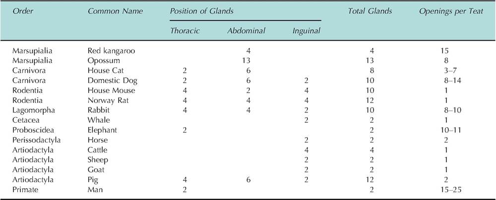
Unlike common dairy species (cows, goats, or sheep), aquatic mammals, especially those in cold environments, produce milk very high in lipid content with little or no lactose. Milk fat provides the suckling young the opportunity to rapidly produce a layer of insulating fat for protection from the cold and to provide a source of metabolically derived water. This illustrates the relevance of lactation to provide a strategy for survival of offspring and reproductive success for mammals in multiple niches.For the placental mammals, the number of mammary glands varies markedly between classes and species. However, among those studied (only about 10% of all mammals), each mammary gland has a teat or nipple. For example, lactation is not common in males, but development of small amounts of mammary tissue and limited secretion can occur. Lactation in males has been reported in humans most likely associated with pituitary dysfunction. However, anecdotal tales of "witch's milk" in male and female infants are not rare. Apparently, normal lactation has been reported in male wild fruit bats. Likely, the first reported lactation in a male ruminant was when Aristotle noted in his Historia Animalium that "a he-goat was milked by his dugs (teats) to such effect that cheese was made of the product." These examples illustrate how little is known about mammary development and lactation in many mammals (Table 18.1).
Herds of Holstein cows routinely average 305- day yields of 13,000 kg of milk, and individual cows produce much more milk. Such prodigious production of milk requires massive mammary glands and careful attention to the feeding and management of these impressive animals. Regardless of the rate of milk production, all milk is produced by the epithelial cells of the mammary alveolus. Consequently, to appreciate milk secretion, it is essential to understand the structure and function of the mammary gland. We'll begin by describing the ontogeny of mammary development followed by a description of physiological processes that allow final structural development of the mammary alveoli and onset of functional differentiation of the secretory epithelial cells.
Table 18.1. Variation in location, number, and nipple openings of mammary glands of selected species.