Introduction
The oral cavity is the entry point of the gastrointestinal tract (GIT). It is where digestion begins with mastication of ingesta, which is mixed with salivary enzymes and passed through the oropharynx to the esophagus.
The oral cavity is sampled most commonly due to the presence of ulcerative lesions and/or masses. Sampling techniques, including brushings, aspirates, and impression smears, similar to those utilized elsewhere, can be employed. Care should be taken to ensure sampling at a depth appropriate to the lesion. Superficial sampling, particularly of ulcerated lesions, is often unrewarding.In health, the mouth, including the oropharynx, is lined by stratified squamous epithelium, which is fully keratinized on the tongue, hard palate, and cheeks and may be in various stages of keratinization elsewhere in the oral cavity. The feline tongue is covered with numerous papillae, which are composed of abundant amounts of connective tissue and keratin spines (Figure 6.1). The type and distribution of these papillae vary with life stage and potentially with environment, as papillae are believed to change with feeding behaviors (Haddad, 2019). Food is moistened by salivary secretions that come from numerous salivary glands, which produce mucous and serous secretions. Carnivores, including cats, are unique in that their saliva contains minimal α-amylase, which, in non-carnivores, initiates carbohydrate digestion in the oral cavity (Gelberg, 2021). Histologically, the mouth lacks a muscularis mucosae, submucosa, and muscularis externa, which are present in other sections of the GIT (Figure 6.2; Bacha & Bacha, 2012).
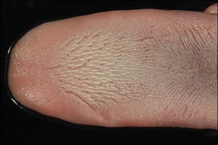
Figure 6.1 Feline tongue with keratin spines. (Courtesy Dr. Raquel Rech.)
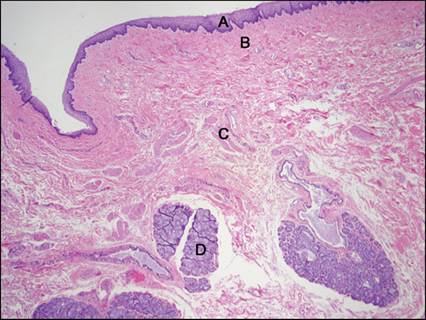
Figure 6.2 Histologic section of the oropharynx of a dog. Stratified, squamous epithelium, keratinized (A); lamina propria (B); skeletal muscle (C); mixed (mucous and serous) salivary gland (D) (H&E, 40? magnification).