‘Normal’ findings
Mixed populations of bacterial organisms, including Actinomyces spp., Fusobacterium spp., and spirochetes, normally inhabit the oral cavity and serve as a defense mechanism, occupying entry/attachment sites of potential pathogens.
Conchiformibius (formerly Simonsiella) spp. are morphologically distinct bacteria that normally populate the oral cavity of warm-blooded vertebrates (Figure 6.3). Conchiformibius spp. form large aggregates that consist of individual organisms (approximately 1.5–6.5 μm long) that ‘slide’ along filaments composed of eight or more cells. Cells on the ends of the filaments can be curved, resulting in a crescent-shaped appearance. Occasionally ‘super filaments’ greater than 50 μm in length result when multiple cells remain attached (Figure 6.4). The presence of Conchiformibius spp. in cytologic specimens is indicative of sampling of or contamination from the oropharyngeal region (Hostetter, 2023). 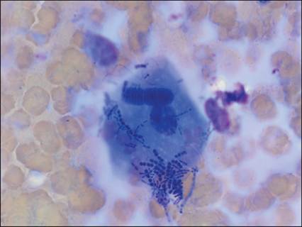
Figure 6.3 Oral cavity from a dog. Keratinizing squamous epithelial cell with adherent mixed bacteria, including Conchiformibius (formerly Simonsiella) spp. (modified Wright’s, 1,000? magnification).
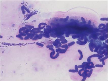
Figure 6.4 Oral cavity from a dog. Conchiformibius (formerly Simonsiella) spp. bacteria, including occasional ‘superfilaments’ (>50 µm long). A mixed population of bacterial rods and cocci is present in the background (modified Wright’s, 1,000? magnification).
In addition to indigenous aerobic and anaerobic bacterial flora, saliva serves as a barrier to pathogenic bacterial colonization, since it can flush microorganisms from the oral cavity and it contains antibacterial compounds (e.g., hydrogen peroxide and thiocyanate), numerous antimicrobial enzymes, and immunoglobulin A (Gelberg, 2021).
Salivary glands, which arise from oral ectoderm, are located throughout the head and neck regions and drain into the oral cavity. In addition to the parotid, mandibular, and sublingual salivary glands found in all species, carnivores have an additional zygomatic salivary gland. Numerous small salivary glands, primarily named by their location (e.g. buccal, labial, lingual, palatine), are present throughout the oral cavity and can exhibit pathology (see Case 1; Gelberg, 2021). Salivary glands are often sampled accidentally with attempted aspiration of submandibular lymph nodes.Cytologic samples from salivary glands are typified by a thick background of stippled eosinophilic mucus, which often exhibits ‘windrowing’, where cells are aligned linearly (Figure 6.5). This pattern, also commonly observed with synovial fluid samples, results from preparation of the thick, viscous specimen. Salivary glands are composed of secretory epithelial cells. Aspirates typically yield cohesive clusters of foamy epithelial cells, often surrounded by more basilar epithelial cells with a high nuclear to cytoplasmic (N:C) ratio (Figure 6.6). Occasionally, amorphous, basophilic mucus droplets are noted both free and within the cytoplasm of the secretory epithelial cells (Figure 6.7).
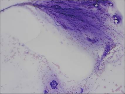
Figure 6.5 Salivary gland from a dog. Basophilic saliva strands admixed with blood in a linear (‘windrowed’) pattern. Small clusters of cohesive, vacuolated salivary epithelial cells (modified Wright’s, 200? magnification).
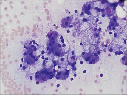
Figure 6.6 Salivary gland from a dog. Clusters of cohesive, uniform, round to polygonal epithelial cells with an abundant amount of basophilic cytoplasm. Most cells contain secretory product, appearing as nonstaining vacuoles. The less vacuolated cells are basal reserve cells (modified Wright’s, 500? magnification).
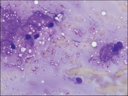
Figure 6.7 Salivary gland from a dog. Individualized salivary epithelial cells releasing amorphous, basophilic mucus globules, which are present both within the cytoplasm and free in the background (modified Wright’s, 1,000? magnification).
Lymphoid tissue in carnivores is found bilaterally in crypts in the dorsolateral aspect of the oropharynx, referred to as palatine tonsils. Dogs and cats typically possess only palatine tonsils, unlike other species that often have tonsillar tissue adjacent to the tongue. Tonsils are covered by stratified squamous epithelium and contain a heterogeneous lymphoid population composed primarily of small lymphocytes with fewer medium and large lymphocytes. Small amounts of extracellular iron are commonly seen (Figure 6.8). Care must be taken not to confuse the presence of well-differentiated squamous epithelial cells in various stages of keratinization from tonsillar aspirates with neoplastic squamous cells, because the tonsils are covered by squamous epithelial cells (Figure 6.9). Unlike other lymphoid tissues, tonsils lack afferent lymphatic vessels and, therefore, do not drain the oral cavity. Their exact role in the immune process is uncertain, and it is speculated that they serve a role in lymphocyte production, antibody formation, and in the innate immune system, as sites of antigen sampling (Weise et al., 2002; Gelberg, 2021).
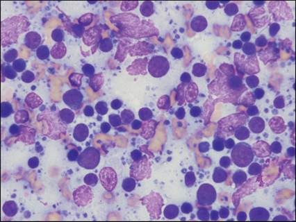
Figure 6.8 Tonsil from a dog. A heterogeneous lymphoid population composed of small, medium, and large lymphocytes. Numerous broken cells and resultant free nuclei are present, along with abundant lymphoglandular bodies (cytoplasmic fragments) (modified Wright’s, 1,000? magnification).
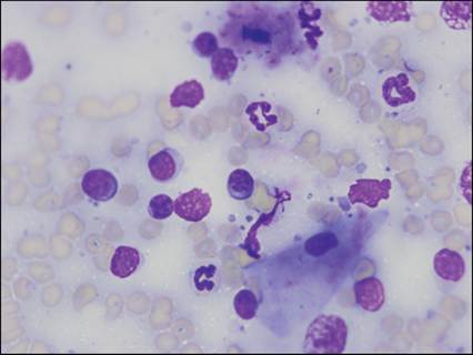
Figure 6.9 Tonsil from a dog. A heterogeneous lymphoid population with blood, lymphoglandular bodies (cytoplasmic fragments), and nuclear material in the background. Extracellular bacteria, including Conchiformibius spp., are noted. A single, keratinizing, nucleated squamous epithelial cell is noted (modified Wright’s, 1,000? magnification).