Inflammation
Neutrophilic inflammation of the oral cavity is often associated with the gingiva (gingivitis), tongue (glossitis), pharynx (pharyngitis), or tonsils (tonsillitis; Figure 6.10). The term ‘stomatitis’ (from the Greek ‘stoma’, meaning opening) is a general term for inflammation within the mouth.
The presence of neutrophils in cytologic specimens from the mouth can be difficult to interpret, because neutrophils at the end of their lifespan are removed via migration through the GIT, including the mouth (Gelberg, 2021). Additionally, as with any cytologic specimen, the contribution of neutrophils from the peripheral blood must be considered. Inflammation can arise secondary to numerous causes, including the presence of bacterial, fungal, and protozoal pathogens and/or foreign material, the latter of which may have a concurrent granulomatous inflammatory response (Figures 6.11–6.14) (Bochnakian et al., 2018). 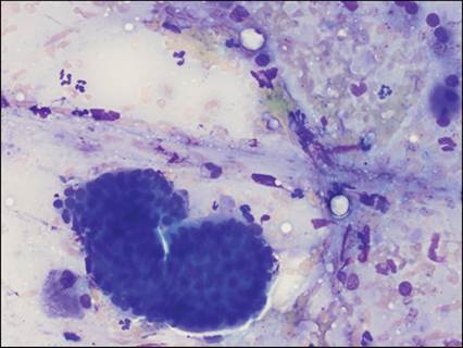
Figure 6.10 Salivary gland from a dog. Cluster of basilar salivary epithelial cells with scattered neutrophils, increased above the amount of blood contamination (modified Wright’s, 500? magnification).
Eosinophilic inflammation is also nonspecific and can be seen with numerous etiologies, including hypersensitivity reactions, parasites, fungal infections, secondary to neoplasia (paraneoplastic), and foreign bodies. Most commonly in cats, but also reported in dogs, are lesions of the ‘eosinophilic granuloma complex’ (Figure 6.15). These lesions, historically termed ‘rodent ulcers’, are of unknown etiology, but may be linked to immune-mediated hypersensitivity. Free, fibrillar collagen strands are often noted within these lesions, due to the presence of collagenolytic factors within eosinophil granules (Figure 6.16; Gelberg, 2021).
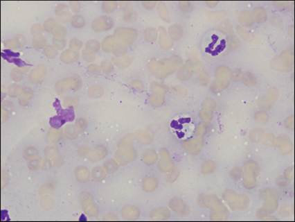
Figure 6.11 Laryngeal polyp from a dog. Thick, basophilic proteinaceous background with blood and broken cells. Two intact segmented neutrophils. Central neutrophil contains small intracellular bacterial cocci (modified Wright’s, 1,000? magnification).
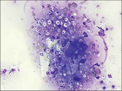
Figure 6.12 Tongue mass from a cat. Keratinized, anucleate squamous epithelial cells with scattered nondegenerate neutrophils and numerous, variably sized basophilic fungal organisms surrounded by a thick, nonstaining capsule, morphologically most compatible with Cryptococcus spp. Culture confirmed Cryptococcus neoformans (modified Wright’s, 500? magnification).
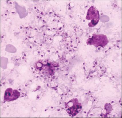
Figure 6.13 Oral plaque from a cat. Numerous 2–4 μm diameter ovoid yeasts with a basophilic, polar nucleus and a clear halo, morphologically compatible with Histoplasma capsulatum. Scattered disrupted inflammatory cells are present. This organism can occasionally be found in tissue drainage and morphologically resembles Sporthrix schecknii, from which it must be distinguished (Wright–Giemsa, 1,000? magnification).
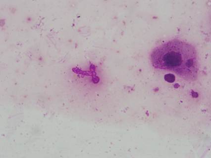
Figure 6.14 Oral plaque from a dog. Fungal pseudohyphal fragment with roughly right-angle branching and bulbous ends, morphologically most compatible with Candida spp. A single nucleated, angular squamous epithelial cell is present (modified Wright’s, 1,000? magnification).
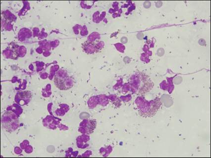
Figure 6.15 Ulcerated lip lesion in a cat. Numerous intact and broken eosinophils with fewer neutrophils.
Abundant rice-grain shaped free eosinophil granules are noted in the background (modified Wright’s, 1,000? magnification).
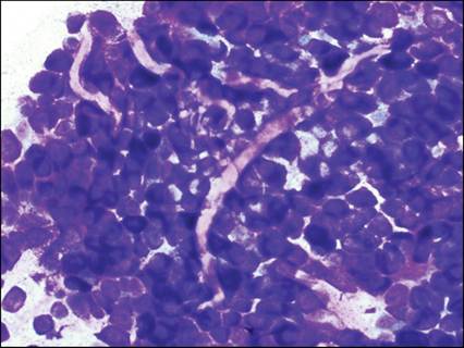
Figure 6.16 Mass aspirate from a cat. Irregularly branching bands of brightly eosinophilic to nonstaining collagen coursing through heavily granulated mast cells (mast cell tumor) (modified Wright’s, 500? magnification).
Lymphocytic, granulomatous, and mixed inflammation can also occur within the oral cavity and have similar significance as elsewhere in the body. Mixed inflammation is often seen with calcinosis circumscripta (dystrophic mineralization), which occasionally presents as hard raised lesions under the tongue (Figure 6.17). Specific causes of this lesion are diverse and include trauma and the presence of foreign material (e.g. suture material) (Gelberg, 2021).
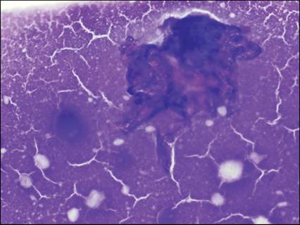
Figure 6.17 Tongue from a dog. Calcinosis circumscripta. Amorphous basophilic mineralized material and a single, poorly distinguishable aggregate of presumed macrophages (Diff-Quik®, 500? magnification).