Introduction
The external ear functions as a conduit for sound to the middle ear and is also important for expression of behavior. The external ear is composed of the pinna (or auricle), the external auditory meatus (the entrance to the ear canal), and the cartilaginous ear canal.
There is a small osseous component to the ear canal of the dog and cat, which is more developed in large animals such as horses and cattle. The external ear canal ends at the tympanic membrane (ear drum).The convex (outer) aspect of the pinna consists of a covering of haired skin comprising epidermis, dermis, subcutis (fat, lymphatics, vessels, and nerves), and skeletal muscles, which overlie the auricular cartilage. The cartilage is elastic and contains many foramina through which blood vessels and nerves traverse (Calhoun & Stinson, 1987; Evans, 1993; Cole, 2009). The concave (inner) aspect of the pinna is more sparsely haired but also comprises epidermis, dermis, and a subcutis that is attached to the concave aspect of the auricular cartilage.
The ear canal is lined by stratified squamous epithelium, which is typically 3–5 cells thick. Variable numbers of hair follicles are present in the ear canal, the quantities of which are highly breed dependent, especially in dogs. The superficial dermis contains sebaceous glands, which open directly into hair follicles. There are also specialized, modified apocrine sweat glands (ceruminous glands) that open either into hair follicles or directly onto the surface of the epidermis. The density of ceruminous and sebaceous glands within the dermis also varies between breeds (Cole, 2009). Deeper in the dermis, there are scattered lymphoid follicles, which are important for local immunity. There are also nerves and vessels throughout the dermis and subcutis. The deepest tissues are the structural components of cartilage and trabecular bone (Figure 18.1).
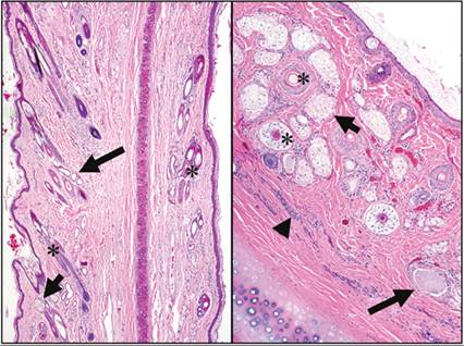
Figure 18.1 Sections of pinna and ear canal; normal structures. Left: section of normal pinna from a dog. The auricular cartilage is in the center and is covered by haired skin consisting of a thin epidermis of stratified squamous epithelium, a dermis with hair follicles (asterisks), sebaceous glands (short arrow) and ceruminous glands (long arrow), and a subcutis. Note that the hair follicles are more numerous on the convex surface (left side). Right: ear canal from a dog. This tissue shows mild hyperplastic changes (mild epidermal hyperplasia, mild dermal fibrosis) and has mild multifocal lymphoplasmacytic infiltrates (arrowhead). Note the hair follicles (asterisks), sebaceous glands (short arrow), ceruminous glands filled with inspissated secretory material (long arrow), and the structural cartilage to the lower right (H&E: left, 40? magnification; right, 100? magnification).
As well as the dermal lymphoid tissue, the ceruminous material that resides within the ear canal is an important component of aural immunity and defense. Cerumen is acidic and composed of sloughed anucleate keratinocytes, watery ceruminous and mixed lipid sebaceous gland secretions, as well as immunoglobulins, lysozyme, and interleukins (Huang et al., 1994; Cole, 2009; Crain et al., 2009). The self-cleaning function of the external ear canal is an effective means of regulating the ceruminous material and is highly important for aural health. Sloughed anucleate keratinocytes migrate to the periphery of the tympanic membrane and then, along with glandular secretions and resident yeasts and bacteria, move outwards to the external auditory meatus to be shaken, scratched, or groomed out of the ear canal (Cole, 2009). Overzealous use of ceruminolytics and ear cleaners impairs this normal process (Rosychuk, 1994).
Cytologic examination of the ear canal is an important part of the diagnostic work up of otic disease in cats and dogs.
Specifics of sampling this region will be discussed further below. All descriptions forthwith refer to staining characteristics with Romanowsky-type stains such as Diff-Quik® (modified rapid Romanowsky stain) or Wright–Giemsa.In healthy dogs and cats, the normal external canal may contain yeasts, cocci, and keratinocytes. The most common yeast in the external ear canal of cats and dogs is Malassezia pachydermatis. The yeasts are peanut-shaped, dark basophilic, and approximately 3 μm wide and 7 μm long (Figure 18.2). There may also be 1 μm diameter dark basophilic cocci present singly, in pairs, or in small clusters. The vast majority of keratinocytes are anucleate (squames), but rare nucleated keratinocytes, also known as squamous epithelial cells, may be seen. Squames are large, angular, and flat or rolled up (‘keratin bars’), and variably stain clear to pale basophilic to eosinophilic. They may contain variable numbers of very fine, bright pink to purple keratohyalin granules. Keratohyalin granules may also be free in the background. Care must be taken not to confuse these granules with cocci – bacteria are larger and typically stain a darker purple/blue (Angus, 2004). Depending on the pigmentation of the dog or cat, scattered coarse granules of brown to gold to black melanin pigment may be seen within squames or free in the background. Cerumen is lipid-rich, and therefore resists staining. It may be seen as amorphous clear material in the background, and may appear to dissolve or disappear with the addition of immersion oil (Figure 18.3).
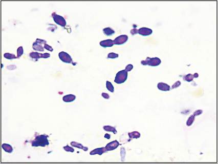
Figure 18.2 Ear swab from a cat; Malassezia pachydermatis yeast. Note the classic peanut or shoe print appearance of the dividing cells. The yeasts are approximately 3 ? 7 μm in size and exhibit unipolar budding on a broad base forming a prominent collarette (modified Wright–Giemsa, 2,000? magnification).
(Courtesy Dr. Kelli Ferris.)
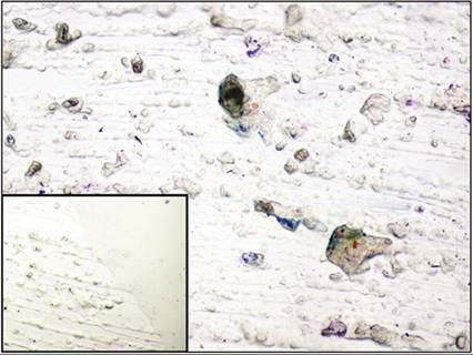
Figure 18.3 Normal ear swab from a cat; cerumen and keratin. Refractile, non- to poorly staining cerumen is admixed with angular squamous cells. Inset: immersion oil has been added to the slide and is dissolving the lipid-rich cerumen (modified Wright–Giemsa, 40? magnification; inset: 100? magnification). (Courtesy Dr. Kelli Ferris.)
Tater et al. (2003) have evaluated the cytology of the vertical ear canal of normal dogs and cats (Table 18.1). Roll preparation cytologic smears were found to be the most cellular. Dogs (n = 50) were found to have a median of 0.2 Malassezia spp. yeast and 3.9 keratinocytes/high-power field (hpf) (400? magnification). In addition, 42% of the dogs also had rare cocci, but the median number was 0/hpf. In cats (n = 52), approximately 8 keratinocytes/hpf were observed and there were 0.2/hpf of Malassezia yeast and 0.3/hpf of cocci. Rod-shaped bacteria were not seen, and are not considered a normal finding in the ears of healthy cats or dogs (Tater et al., 2003). The presence of neutrophils within the ear canal is always abnormal and indicates inflammation (Ginel et al., 2002).
Table 18.1 Cytologic findings of the external ear canal in the normal dog and cat
| Median number per high-power field (400? magnification) | ||
| Feature | Dog | Cat |
| Malassezia yeast | 0.2 | 0.2 |
| Cocci | 0 (rare) | 0.3 |
| Keratinocytes | 3.9 | 8 |