Sampling
Cytologic examination is warranted for any lesion of the pinna or external ear canal. Some lesions may exfoliate poorly, such as mesenchymal tumors (e.g., fibrosarcoma, hemangioma, hemangiosarcoma).
Nodules can be aspirated either with or without negative pressure from an attached syringe. Swabbing ear canal nodules is unlikely to reveal the cause. Masses within the ear canal are best aspirated in a heavily sedated or anesthetized animal, facilitated via an otoscope or an otoendoscope. The aspirated material should be rapidly discharged and gently smeared across one or multiple slides. Fine needle aspiration (FNA) of the regional submandibular or retropharyngeal lymph nodes may also be indicated.On the pinna, flat lesions may be best assessed by impression smears of any exudative or eroded/ulcerated surface, followed by careful scraping of the underlying lesion with a spatula or a dulled scalpel blade. The material may then be wiped across multiple slides. This is the most useful way of cytologically diagnosing squamous cell carcinoma (SCC).
Alopecic or crusted lesions can be assessed with hair plucks for dermatophytes or Demodex mites (Figure 18.4), and/or multiple superficial and deep skin scrapings in mineral oil for Sarcoptes or Demodex mites, respectively (Figure 18.5). For ceruminous debris on the pinna, a dulled blade may be used to gently collect some of the material, which is then wiped across slides without oil for staining. Organisms such as M. pachydermitis or Leishmania spp., as well as bacteria, may be identified in this way. For drier, less exudative lesions of the pinnae, tape strip sample collection is useful and less traumatic to the inflamed skin (Figure 18.6).
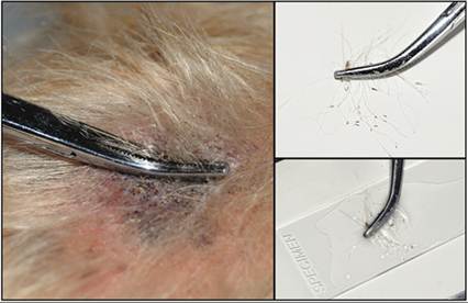
Figure 18.4 Hair pluck preparation from a dog with suspected demodicosis.
Left: an area with evidence of follicular pathology (broken hairs, comedones, etc.) is selected. A sample of hair is firmly grasped with forceps, close to the skin, and is quickly plucked. Right top: a good quality sample will have a sufficient number of hairs, including the roots, to allow thorough examination. Bottom right: mineral oil is placed on a glass slide and the hairs are added. A coverslip will be placed on top to allow easy viewing with a microscope. The slide should be examined on both low-power (40?/100?) and high-power (400?) magnification.
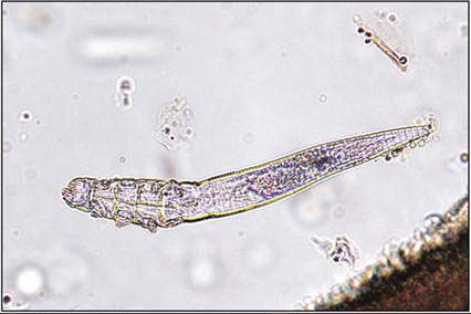
Figure 18.5 Deep skin scraping from a cat; demodicosis. The preparation shows the typical, elongated, ‘cigar-shaped’ appearance of Demodex cati. Adult mites range in length from approximately 180 to 220 μm, have a long and prominently striped abdomen, and four sets of clawed appendages. Because this mite inhabits hair follicles, hair plucks or deep scrapings are the best methods to use for screening (unstained oil mount preparation, 200? magnification).
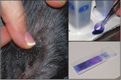
Figure 18.6 Tape strip sample preparation from the pinna of a dog. Left: tape is firmly pressed onto the lesion. Tape strip preparations are best used with drier, less exudative skin lesions. Top right: the tape strip is stained using only the third, dark blue solution of the quick stain and then briefly rinsed in water. Bottom right: the stained preparation is placed on a slide, specimen side down, and then blotted with blotting paper to removed excess liquid.
Cytologic examination of ceruminous and/or purulent debris is indicated for every case of otitis externa. It is also useful as a tool for monitoring response to treatment. Cytology is considered more sensitive for detecting Malassezia yeast and bacteria than culture (Huang et al., 1994; Griffin et al., 2007).
Although sampling the horizontal ear canal is likely more representative of the infection, it can be difficult in an awake animal and sedation or anesthesia is not necessarily indicated for every case of otitis externa. In most cases, sampling the junction of the vertical and horizontal canals is sufficient (Angus, 2004). To optimize the sampling of the external ear canal, gently pull up on the pinna to help straighten the ear canal and then place the swab into the ear canal, angling toward the nose, and slowly advance. The swab should be rolled into the debris and then removed from the ear and rolled across glass slides. For collection of samples to search for ear mites such as Otodectes cynotis or Demodex spp., particularly in cats, a drop or two of mineral oil is placed on a separate glass microscope slide and the same swab used to collect the cytology sample is dipped into the mineral oil. The swab is then gently placed back into the ear canal and debris collected. The debris is then transferred to the same glass microscope slide containing the mineral oil by gently rolling the swab in the drop of mineral oil. Both ears should be sampled with separate swabs on separate slides, and the slides carefully labeled. Slides should be examined promptly since mites may possibly walk off the slide if left for too long.For optimal cytologic examination, preparation of multiple slides is recommended when collecting samples from nodules or masses of the pinna or ear canal. In addition, it is always useful to keep at least one slide unstained from any type of otic cytology in case special stains are required.
Skin scrapings prepared with mineral oil are cover slipped and examined under the microscope directly. Hair plucks may be cleared with potassium hydroxide to search for dermatophytes. Other preparations should be stained. Heat fixation of the slide does not improve staining; thus rapid air-drying of smears is adequate for cytology (Toma et al., 2006; Griffin et al., 2007). Romanowsky-type stains such as Diff-Quik®, or similar modified rapid stains, or Wright–Giemsa are the most useful. The lipid component of cerumen may be dissolved by the alcohol in the staining process (Ginel et al., 2002).
It is desirable to possess a separate set of jars of rapid stain dedicated for suspected infectious or so called ‘dirty’ ear cytology specimens to prevent contamination of other samples. Cytology of otic debris can be stained in the usual fashion, or, for more rapid results, a single 5–10 second dip in the counterstain (purple stain) is considered sufficient for identifying bacteria, yeast, keratinocytes, and neutrophils (Toma et al., 2006). For tape preparations, jar 1 should not be used as the alcohol will remove the glue component from the tape and thus your sample.