Laboratory evaluation
The clinician should measure and report the amount of fluid removed from the body cavity and comment on possible iatrogenic hemorrhage. Gross examination evaluates color, turbidity, odor, and viscosity.
This should be done by the clinician when the sample is submitted, because color might change during storage and transport. Gross examination must not be neglected as it often yields valuable information regarding the underlying disease (Figures 15.6, 15.10, 15.11, 15.16, 15.17, 15.21, 15.22, 15.52–15.57).Physiologic fluids and/or transudates are usually clear, colorless to slightly yellowish fluids with a low viscosity and without a discerning odor (Figure 15.52). Blood (Figures 15.10, 15.11), chylous lymph (Figure 15.16), bile, urine, and septic and nonseptic inflammation can cause a multitude of color changes. All shades of red to pink signal hemorrhage. As long as the red blood cells (RBCs) remain intact, the supernatant is clear; in cases of hemolysis the supernatant will be reddish. Beige to creamy or reddish-brown coloration is often seen in septic exudates (Figures 15.53–15.55).
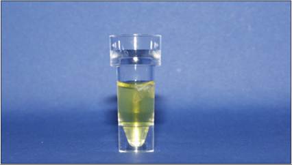
Figure 15.52 Transudate from a dog. The fluid is pale yellow and clear.
Turbidity is caused by light diffraction, which can be elicited by particles, such as cells (Figures 15.53, 15.54), or cholesterol-containing lipids or chylous fluid. The presence of chylomicrons causes a milky white opaque appearance (Figures 15.16, 15.17). When chylous fluid is allowed to stand, a creamy layer is formed on top (Figure 15.17). Turbid samples with high cellularity clear when cells follow gravity and sink or are spun down by centrifugation. Turbidity caused by cholesterol-rich lipids does not clear when allowed to stand or centrifuged.
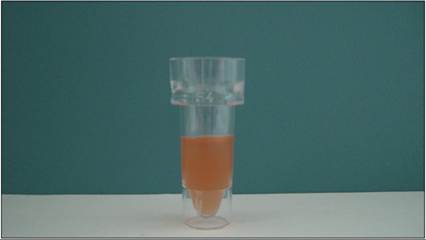
Figure 15.53 Effusion from a dog.
This fluid is an exudate. Exudates have a high cell count and protein concentration and are therefore often turbid.
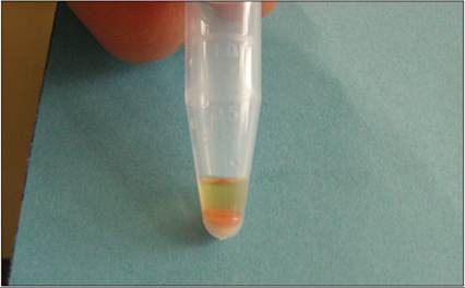
Figure 15.54 Effusion from a dog. The sample was centrifuged. Note the thick cell pellet post-centrifugation.
Viscosity is due to the friction between neighboring particles in a fluid, which are moving at different velocities. Viscosity in effusions can change due to an increase in inflammatory proteins or increased cellularity. FIP-associated exudates are typical examples (Figure 15.6). Coagulated material might cause a flocculent appearance. ‘Sulfur granules’ (yellow grains in reddish brown turbid exudates) are a typical finding in septic exudates caused by Nocardia or Actinomyces spp. (Figures 15.56, 15.57). A foul odor can also occur with septic inflammation.
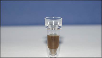
Figure 15.55 Effusion from a dog. This patient had a penetrating foreign body. The sample is markedly turbid and brown.
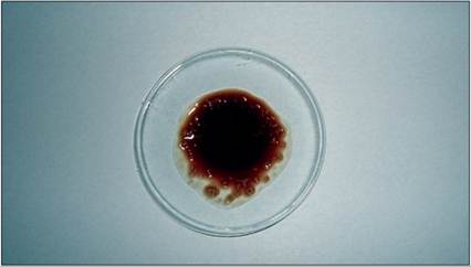
Figure 15.56 Pleural effusion from a cat. This patient had a septic pleuritis with Nocardia spp. Note the sulfur granules.
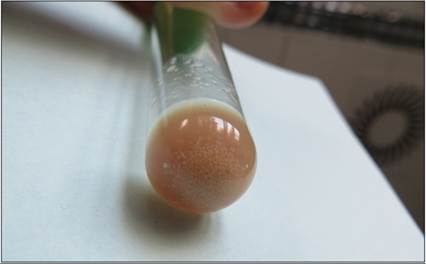
Figure 15.57 Pleural effusion from a cat. This patient had septic pleuritis with Actinomyces spp. Again, note the sulfur granules.
Microbiologic cultures
Fluid samples collected into sterile tubes without anticoagulant (EDTA can be bacteriostatic) may be submitted for bacterial and fungal cultures. The reference laboratory should be contacted for special culture conditions when indicated. Fluids should be cultured for both anaerobic and aerobic bacteria.
Biochemistry analytes
A variety of clinical chemistry analytes are helpful in determining the nature of an effusion.
Measurement of total protein concentration is always performed to differentiate between transudates and other types of effusion. The other biochemistry tests are only required in specific clinical situations. Different brands of benchtop analyzers might vary considerably in their analytical performance when used for the analysis of body cavity effusions (Hetzel et al., 2012). This is not surprising because the expected concentrations of some analytes in body cavity effusions are beyond or close to the analytical range of various assay systems. Therefore, very often numerical results cannot be obtained.Total protein concentration can reliably be determined by refractometry, the biuret method, and urine test strips (Braun et al., 2001; Papasouliotis et al., 2002). The measurement should be made from the supernatant of a centrifuged sample to avoid measurement errors. However, it has to be kept in mind that refractometry measurements for concentrations of 2.5 g/dl (25 g/l) are less accurate than photometric testing by the biuret method. The protein test pad on commercial urine test strips can also be used for a semi-quantitative protein assessment (Braun et al., 2001). Effusions with a protein concentration of 2.5 g/dl (25 g/l) are categorized as transudates; when the protein concentration lies between 2.5 and 3.5 g/dl (25 and 35 g/l) and cellularity is low, the effusion is categorized as a protein-rich transudate (previously described as modified transudate), whereas effusions with high cellularity and protein concentration are exudates.
Triglyceride concentration is measured to confirm the nature of a chylous effusion. A concentration of >100 mg/dl (11.3 mmol/l) is considered diagnostic. However, in anorectic animals, the concentration might be lower. In those cases, comparison with blood triglyceride concentration might be helpful (Dempsey & Ewing, 2011; Valenciano et al., 2013).
Cholesterol concentration in effusions exceeding plasma cholesterol concentration aids the identification of pseudochylous effusions.
Glucose concentration determination and calculating the difference between blood glucose concentration and glucose concentration in the body cavity fluid have been considered helpful in differentiating septic from nonseptic effusions (Bonczynski et al., 2003). A difference in glucose concentration between blood and effusion above 20 mg/dL has been suggested as highly suspect for a septic process (Dempsey & Ewing, 2011). Accordingly, the concentration of fluid lactate and the difference from blood lactate have been used for the same purpose. These data were based on a low case number, and a later study demonstrated them to be unreliable (Szabo et al., 2011). This is not surprising as all cells consume glucose, so highly cellular effusions will always have a decreased glucose concentration. However, glucose/lactate measurements give just a quick orientation and cannot replace cytologic evaluation and culture testing to confirm a septic process.
Creatinine concentrations at least double the plasma creatinine concentration are expected in cases of uroperitoneum. Sample dilution for creatinine measurement might be warranted to obtain a numerical result. More recently a fluid-creatinine to serum-creatinine ratio of ≥1.25 has been suggested to confirm uroabdomen in dogs (Paes et al., 2022).
Bilirubin concentration is measured in effusions to confirm leakage of bile into an effusion; the bilirubin concentration of the fluid in the effusion exceeds the plasma concentration.
Measurement of bile acid concentration in abdominal effusions is useful to diagnose anicteric gall-bladder rupture, when bilirubin concentrations are inconclusive (Pascual et al., 2022).
Lipase activity measurement can be helpful in establishing a diagnosis of pancreatitis-associated effusion; the enzyme activities in the effusion are higher than in plasma (de Arespacochaga et al., 2006).
Lactate dehydrogenase (LDH) activity measurement has been used in humans to differentiate between benign and malignant ascites caused by ovarian tumors and to differentiate between benign and malignant pleural effusions (Halperin et al., 1998; Bielsa et al., 2008).
However, LDH is an enzyme present in most cells, so its activity is correlated with cellularity. LDH measurement is also a component of Light’s criteria, a classifications system established in human medicine to differentiate between exudates and transudates which has been evaluated for veterinary patients (Zoia, Slater et al., 2009; Zoia, Petini et al., 2020; Alonso, Mattoso et al., 2021).Plasma cardiac troponin I was found to be helpful in differentiating dogs with cardiac hemangiosarcoma from dogs with hemangiosarcoma of other sites or other tumors, and dogs with pericardial effusions of other than neoplastic origin (Chun et al., 2010). However, there was an overlap of values between the groups, so this test might be of limited value in the assessment of a single patient, without specific findings of a cardiac mass in diagnostic imaging.
Plasma or effusion fluid N-terminal pro brain natriuretic peptide (NT-proBNP) is useful in identifying congestive processes which contribute to the formation of effusions, especially in cats (Humm et al., 2013; Wurtinger et al., 2017; O'Shaughnessy et al., 2022). In dogs with cardiac tamponade, an increase of NT-proBNP has been observed after removal of the pericardial fluid, probably due to improved ventricular filling pressure (Baumwart et al., 2017).
Acid–base status, electrolytes, glucose, and lactate concentration in blood and pericardial effusion were considered diagnostic in differentiating dogs with neoplastic from those with nonneoplastic pericardial effusions. The overlap between the two groups is wide, so diagnostic use of these tests for categorizing a single patient is fairly limited (de Laforcade et al., 2005).
Under field conditions, measurement of leukocyte esterase in peritoneal fluid of dogs might be useful to rule out bacterial peritonitis, because a negative test result has a high specificity (Thomovsky et al., 2014).
Coagulation testing in peripheral blood is routinely performed to investigate the cause of hemorrhagic effusions.
Measurements of fibrinogen degradation products (FDPs), D-dimer, fibrinogen concentration in effusions and peripheral blood, and viscoelastic measurements in dogs with all types of cavitary effusion indicate enhanced fibrinolytic activity. (Zoia, Drigo et al., 2022). This is caused by proteins involved in hemostasis that spill into cavitary effusions regardless of their mechanism of formation into a milieu in which the actions of these proteins are not well regulated, owing to the lack of other constituents of the hemostatic system such as platelets or vascular endothelium. (Zoia, Drigo et al., 2022).Cell concentrations are usually determined by electronic cell counters or a hemocytometer (Bauer & Moritz, 2005; Pinto da Cunha et al., 2009; Stockham Scott, 2013). Cell clumping, cell fragmentation, and noncellular debris can cause erroneous cell counts (Cowell et al., 1987; Pinto da Cunha et al., 2009). Advanced hematology analyzers might provide additional information regarding the cellular composition, such as indicating tumor cells (Bauer & Moritz, 2005); however, microscopic slide evaluation must not be omitted. Malodorous, flocculent effusions (Figures 15.55–15.57) should not be introduced into an electronic counter, because they might block and contaminate tubing. In these cases, estimation of cellularity by evaluation of an appropriately prepared direct smear suffices.