Macrorhabdus Ornithogaster infection in BIRDS
DESIREE S. JANSSON
Department of Animal Health and Antimicrobial Strategies, National Veterinary Institute & Department of Biomedical Sciences and Veterinary Public Health, Swedish University of Agricultural Sciences, Uppsala, Sweden
Macrorhabdus ornithogaster (formerly known as ‘megabacterium’) is a yeast that colonizes the gastric tissues of some birds.
The disease is called megabacteriosis, ‘going light’, ‘chronic wasting disease’ or ‘thin bird disease’.Macrorhabdus ornithogaster is an ascomycetous yeast and the sole known member of the genus Macrorhabdus(24). The organism is filamentous and 2—3 ? 20—80 μm in size. Cells possess a single nucleus, a deeply invaginated plasma membrane, a thick chitinous cell wall, and septae, but they seem to lack mitochondriae(24-26). Sequence data of partial 18S rRNA genes are available from two budgerigars and show 97% sequence similarity1-24’27). It is assumed that morphologically similar organisms from other species belong to the same or a closely related taxon.
Organisms indistinguishable from M. ornithogaster have been reported from passerine and psittacine companion birds, domestic birds (chickens, turkeys, ostriches, partridges), free-living finches (order Passeriformes, family Fringillidae) and sulphur-crested cockatoos ( Cacatua galer- ita). Transmission occurs orally. Presence of this yeast in faeces indicates a faecal source, but bird-to-bird transmission from mutual feeding during courtship or feeding of hatchlings cannot be excluded. An environmental source has not been identified. The mechanisms of pathogenesis are unknown.
Colonization in domestic and companion birds is associated clinically with weight loss, ill thrift, dehydration, fluffed-up plumage, anorexia or apparent polyphagia, regurgitation and diarrhoea or dry faeces of varying degrees of severity.
Sudden death from proventricular haemorrhage or rupture occurs sporadically. The clinical significance in wild finches remains to be determined. Colonization of the gastrointestinal tract by this yeast in wild finches is often observed concurrently with colibacil- losis or salmonellosis(28) (Jansson et al., unpublished observations).Gross lesions consist of an enlarged and hyperaemic proventriculus with a viscous mucoid luminal cover (Figure 40.1). Microscopically, the organism is visualized by H&E stains (Figure 40.2), which improves with silver and PAS stains, but it stains weakly or variably by Gram stain. Microscopically, lymphoplasmocytic and/or heterophilic inflammation, epithelial hyperplasia, disrupted koilin layer, goblet cell hyperplasia, micro-abscesses and focal necroses may be observed.
Ante mortem diagnosis depends on identification of the organism in faeces or crop/gastric washings. Post mortem diagnosis is achieved by demonstration of the organism in
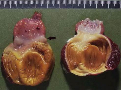
FIGURE 40.1 Hyperaemic proventriculus and mucinous covering of the isthmus region (arrow) in a partridge (Perdix perdiX) colonized by presumed Macrorhabdus ornithogaster (left), and a normal organ (right). Photo: Bengt Ekberg, SVA.
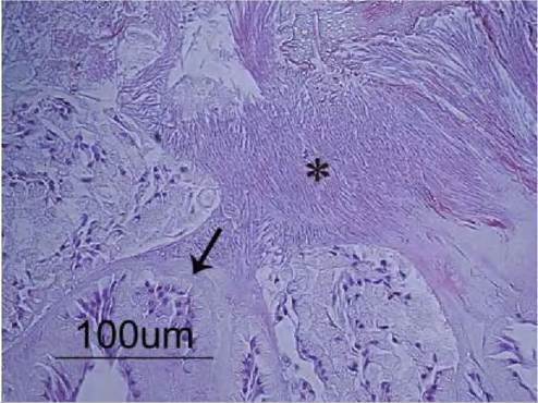
FIGURE 40.2 Macrorhabdus ornithogasterAdke organisms in the isthmus region of a free-living wild common redpoll ( Carduelisflammea) diagnosed with salmonellosis. The organisms display disorganized superficial growth and profuse oriented growth (*) in the mucinous layer that covers the epithelium (arrow). (H&E) Photo: DesireeJansson.
the proventriculus by microscopy. Isolation requires liquid or semi-solid Basal Medium Eagle at pH 3—4 with 20% fetal calf serum, glucose or sucrose as growth substrate, and microaerophilic incubation at 42°C for 3 days(27). Treatment is possible with oral amphotericin B, and can resolve clinical signs but will not always eliminate the organism.
Colonization of mammals has not been confirmed. Macrorhabdus ornithogaster was successfully transferred from budgerigars to chickens(27,29). Hence, natural transmission from companion birds and wildlife to domestic birds should be considered as a potential disease risk.