Geomyces Destructans - WHITE-NOSE SYNDROME IN HIBERNATING BATS
contribute to the spread of G. destructans. There is also evidence of environmental persistence of this organism in cave hibernacula in the absence of bats.
Bats infected with G.
destructans may not have white fungal growth on the muzzle, and the white muzzle is not diagnostic for infection with G. destructans. Wing membrane affected with the G. destructans can adhere to other wing folds and may lead to tearing of the wing membrane as the wings are extended. Microscopically, the characteristic pattern of ‘cupping erosion’ is formed by dense, focal aggregates of fungal hyphae at the interface of the skin(33). As these colonies of fungal hyphae proliferate and expand, they erode the epidermis and eventually invade all layers of skin with ulceration and invasion of deep dermal structures. PAS stain is essential to identify these clusters of fungal hyphae and the distinct pattern of erosion and invasion that is currently the diagnostic feature ofWNS (Figure 40.3). The invasion of living tissue distinguishes G. destructans from typical dermatophytes of mammals. The lack of cellular response to G. destructans as it erodes and invades the skin is a likely consequence of physiological immunosupression in normal hibernating mammals. Recovery from WNS as bats become euthermic has been documented in rehabilitation studies(34). Wing membrane is critical to physiological homeostasis in hibernating bats.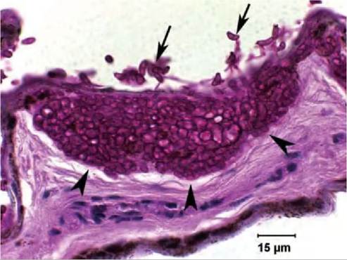
FIGURE 40.3 Wing membrane from M. lucifugus with WNS collected in the USA (PAS stain). Typical cupping erosion of skin at the advancing margin of fungal hyphae (arrowheads). Conidia characteristic of G. destructans are present (arrows). Note the lack of cellular inflammatory response to the fungus. Photo: Carol U. Meteyer, USGS, NWHC.
Damage to the integrity of wing membrane with subsequent disruption of hydration, circulation, cutaneous respiration and thermoregulation is the proposed mechanism by which infection with G. destructans kills hibernating bats(35).
Behavioural signs of bats with WNS include increased frequency and duration of arousal cycles, shifts from normal roost sites deep in the caves towards cave entrances, and early emergence from hibernation with day flights during winter. These abnormal behaviours lead to premature consumption of energy reserves, and can result in emaciation, which is another finding often associated with WNS.
Because G. destructans spores can be present on bats without causing infection, the current confirmatory test for WNS is microscopic identification of the typical histologic skin lesions described above. If the muzzle of a dead bat is not visibly affected, wings are the best tissue for histology, fungal isolation and polymerase chain reaction (PCR). Growth on Sabouraud dextrose or cornmeal agar is optimal, although slow, at 5-10°C. Microscopic identification of G. destructans conidia on fungal tape collections from a clinically affected muzzle can provide a non-lethal screening tool for G. destructans with subsequent confirmation using histopathology, culture or PCR.
Current management strategies are directed at reducing the potential for human spread of G. destructans through decontamination of footwear and equipment, and closing caves and mines to human access. There is no currently accepted treatment for bats that is efficacious, reasonable to administer without disrupting hibernation, a viable option for treating large numbers of bats, and known to be safe. Treatment of hibernacula is currently untenable because of the risk to other cave biota and delicate cave ecosystems. Because of the unique environmental and host physiology requirements for successful pathogenicity of G. destructans, there appears to be little risk of this disease spreading to non-hibernating warm-blooded animals, including humans.
Potential for spread to other hibernating mammals, insects and exotherms is unknown.REFERENCES
1. Rochette, F., Engelen, M. & Vanden Bossche, H. Antifungal agents of use in animal health-practical applications. Journal of Veterinary Pharmacology and Therapeutics. 2003;26:31-53.
2. Fischer, O.A. Adiaspores of Emmonsia parva var crescens in lungs of small rodents in a rural area. Acta Veterinaria (Beograd). 2001;70: 345-52.
3. Stroud, H.K. & Coles, E.M. Blastomycosis in an African lion. Journal of the American Veterinary Medical Association. 1980;177:842-4.
4. Thiel, R.P., Mech, L.D., Ruth, G.R., Archer, J.R. & Kaufman, L. Blastomycosis in wild wolves. Journal of Wildlife Diseases. 1987;23: 321-3.
5. Shubitz, L.F Comparative aspects of coccidioidomycosis in animals and humans. Annals of the New York Academy of Sciences. 2007;1111: 395-403.
6. Burtscher, H. & Otte, E. Histoplasmose beim chinchilla. Deutsche Tierarztliche Wochenschrift. 1962;69:303-7.
7. Farinas, F., Flores, L., Rodriguez, P., Sabalete, T. & Quevedo, M.A. Disseminated histoplasmosis in a dorcas gazelle ( Gazella dorcas neglecta) kept in captivity conditions in Spain. Revista iberoamericana de micologia. 2009;26:152 4.
8. Dei-Cas, E., Chabe, M., Moukhlis, R. et al. Pneumocystis oryctolagi sp. nov., an uncultured fungus causing pneumonia in rabbits at weaning: review of current knowledge, and description of a new taxon on genotypic, phylogenetic and phenotypic bases. FEMS Microbiology Reviews. 2006;30:853-7.
9. Laakkonen, J. Pneumocystis carinii in wildlife. International Journal for Parasitology. 1998;28:241-52.
10. Kaplan, W., Broderson, J.R. & Pacific, J.N. Spontaneous systemic sporotrichosis in nine-banded armadillos (Dasypus novemcinctus) Sab- ouraudia. 1982;20:289-94.
11. Migaki, G., Font, R.L., Kaplan, W & Asper, E.D. Sporotrichosis in a Pacific white-sided dolphin (Lagenorhynchus obliquidens). American Journal of Veterinary Research.
1978;39:1916-9.12. Quesada, O., Rodriguez, F., Herraez, P., Seara, D. & Espinosa de los Monteros, A. Mucor ramosιssιm-us associated with feather loss in canaries (Serinus canarius). Avian Diseases. 2007;51:643-5.
13. Chayakulkeeree, M., Ghannoum, M.A. & Perfect, J.R. Zygomycosis: the re-emerging fungal infection. European Journal of Clinical Microbiology and Infectious Diseases. 2006;25:215-29.
14. French, R.A. & Ashworth, C. D. Zygomycosis caused by Conidiobolus coronatus in a Llama (Lama glamci). Veterinary Pathology. 1994;31: 120-2.
15. Silva, S.M., Castro, R.S., Costa, F.A. et al. Conidiobolomycosis in sheep in Brazil. Veterinary Pathology. 2007;44:314-9.
16. Keymer, I.F., Gibson, E.A. & Reynolds, D.J. Zoonoses and other findings in hedgehogs (Erinaceus europaeus): a survey of mortality and review of the literature. Veterinary Record. 1991;16:245-9.
17. Bexton, S. & Robinson, I. BSAVA manual of wildlife casualties. Mul- lineaux, E., Best, D. & Cooper, J.E. (eds). British Small Animal Veterinary Association, Gloucester; 304 pages, 2003; pp.49-65.
18. Morris, P.A. & English, M.P. Transmission and course ot Trichophyton erinacei infections in British hedgehogs. Sabouraudia. 1973;11:42- 7.
19. Smith, J.M.B. & Marples, M.J. Trichophyton mentagrophytes var erinacei. Sabouraudia. 1963;3:1—10.
20. English, M.P. & Morris, P. A. Trichophyton mentagrophytes var ennncer in hedgehog nests. Sabouraudia. 1969;7:118-21.
21. Morris, P.A. Nightly movements of hedgehogs (Erinaceous europeaut) in forest edge habitat. Mammalia. 1986;50:395-8.
22. Peano, A., Tizzani, P., Gallo, M.G. et al. Dermatophytosis due to Trichophyton verrucosum in a chamois (Rupicapra rupicaprot). European Journal of Wildlife Research. 2008;54:153-6.
23. Gallo, M.G., Tizzani, P., Peano, A., Rambozzi, L. & Meneguz, P.G. Eastern cottontail (Sylvilagus floridanus) as carrier of dermatophyte fungi. Mycopathologia. 2005;160:163-6.
24. Tomaszewski, E.K., Logan, K.S., Snowden, KT., Kuzman, C.P. & Phalen, D.N. Phylogenetic analysis identifies the ‘megabacterium’ of birds as a novel anamorphic ascomycetous yeast, Macrorhabdus orni- thogaster gen. nov., sp. nov. International Journal of Systematic and Evolutionary Microbiology. 2003;53:1201-5.
25. Ravelhofer-Rotheneder, K.H., Engelhardt, O., Wolf, R., Amann, W, Breuer, W. & Kosters, J. Taxonomische Klassifizierung von “Megabakterien”-isolaten aus Wellensittichen (Melopsittacus undulatus Shaw, 1805). Tierarztliche Praxis. 2000;28:415-20.
26. Jansson, D.S., Brojer, C., Mattsson, R., Feinstein, R., Morner, T & Hard af Segerstad, C. Mycotic proventriculitis in gray partridges (Perdix perdix) in two game bird farms. Journal of Zoo and Wildlife Medicine. 2008;9:428-37.
27. Hannafusa, Y., Bradley, A., Tomaszewski, E.E., Libal, M.C. & Phalen, D.N. Growth and metabolic characterization of Macrorhabdus orni- thogaster. Journal of Veterinary Diagnostic Investigation. 2007;19: 256-65.
28. Pennycott, TW, Ross, H.M., McLaren, I.M., Park, A., Hopkins, G.F. & Foster, G. Causes of death of wild birds of the family Fringillidae in Britain. Veterinary Record. 1998;143:155-8.
29. Phalen, D.N. & Moore, R.P. Experimental infection of white-leghorn cockerels with Macrorhabdus ornithogaster (megabacterium). Avian Diseases. 2003;47:254-60.
30. Blehert, D.S., Hicks, A.C., Behr, M. et al. Bat white-nose syndrome: an emerging fungal pathogen? Science. 2009;232:227.
31. Wibbelt, G., Kurth, A., Hellmann, D. et al. White-nose syndrome fungus ( Geomyces destructans) in bats, Europe. Emerging Infectious Diseases. 2010;16:1237-43.
32. Puechmaille, S.J., Wibbelt, G., Korn, V. et al. Pan-European distribution of white-nose syndrome fungus ( Geomyces destructans) not associated with mass mortality. PLoS ONE. 2011;4:e19167.
33. Meteyer, C.U., Buckles, E.L., Blehert, D.S. et al.. Histopathologic criteria to confirm white-nose syndrome in bats. Journal of Veterinary Diagnostic Investigation. 2009;21:411-4.
34. Meteyer, C.U., Velent, M., Kashmer, J. et al. Recovery of little brown bats (Myotis lucifugus) from natural infection with Geomyces destructans, white-nose syndrome. Journal of Wildlife Diseases. 2011;47:618-26.
35. Cryan, PM. Meteyer, C.U., Boyles, J.G. & Blehert, D.S. Wing pathology of white-nose syndrome in bats suggests life-threatening disruption of physiology. BioMed Central Biology. 2010;8:135.