Main features
Bone structure
To lighten the skeleton the cortex of the bone is much thinner than in mammalian bones and many long bones are hollow. Internally, lightweight struts create a honeycombed interior and reinforce the bones (Fig.
11.2).Many of the larger bones are pneumatic, i.e. they arc filled with air contained in membranous air sacs which connect with the respiratory system. These cut down the weight of the skeleton and so aid flight. The number of pneumatic bones is reduced in diving birds as they restrict the ability to stav under waler.
Skeletal modifications
The sternum is extended into a laterally flattened keel (Fig. 13.1) which provides a large surface area for the attachment of the major flight muscles. These are Iheprctond muscles responsible for the powerful down stroke and the Supracoracoid muscle responsible for the upstroke. Flightless birds, e.g. ostriches and emus, do not have a keel.
To counteract the action of the flight muscles and to support the wings a large bone, the conicoid, lies between the keel and each shoulder joint (Fig. 13.1).
The number of joints in the body is reduced. This is particularly seen in the vertebral column and results in a rigid trunk to support the action of the flight muscles.
The neck is long and mobile and contains more cervical vertebrae than the seven seen in all mammals - there may be as many as 2 5 in the swan. A long flexible neck enables a bird to turn its head, creating the wide range of vision necessary for survival, to catch Hxxi and to preen all parts of its body.
The number of caudal vertebrae are reduced and fused forming the toil or pygostyle. Some flight feathers are attached to the tail and play an important part in flight, balance and display. At the base of the tail is the uropyιjial or preen gland which is essential for keeping feathers oiled and healthy.
V
There is a single hx⅛ cavity, i.e. birds have no diaphragm dividing the thorax and abdomen. There is no floor to the pelvic cavity, which makes the pelvis more distensible to facilitate the passage of eggs to the outside. Egg laying enables birds to reproduce without the need to carry the developing foetus around internally. This would increase the Ixxiy weight and severely restrict the ability to fly.
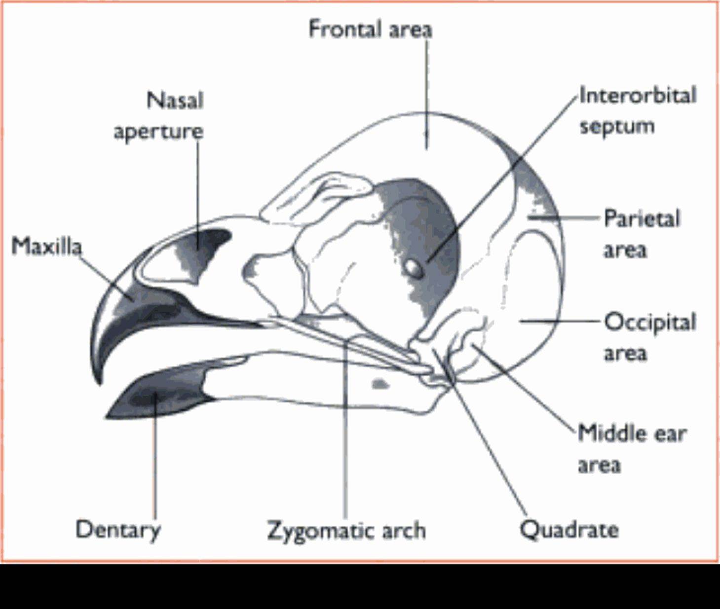
Head (Fig. 13.3)
A lightweight beak covering the mandible replaces the teeth and reduces the weight of the skeleton. Each species has a characteristic beak shape, which is adapted to its eating habits.
Between the mandible and skull is Ihequadrate bone, which makes dislocation of the jaw very unlikely.
Many birds also have a joint known as the Criiniofiiciii! hinge between the upper beak and the skull. This increases the mobility of the beak during feeding.
The orbit is large and thin-walled to lighten the skull and to house the large eyes. As much of the

Fig. 13.1 Skeleton of a typical bird (hawk) (Reprmted from C∣∣∩κal Anatomy and Phys∣ology for Vetermary Technicians1T Colville and JM Bassett, p 355. Copynght 2002. with permission from Elsevier Science.)
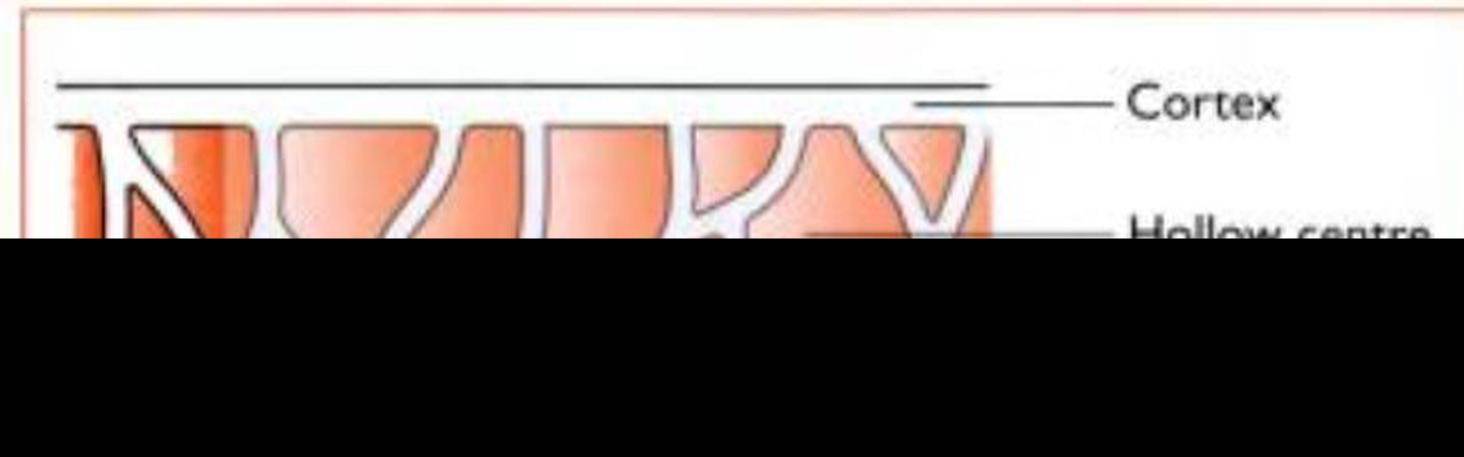
Fig. 13.2 Longitudinal section through an avun >ong bone ' he bony struts serve to strengthen the bone
bird s brain is concerned with visual information, the eyes connect with large optic Iohes in (he brain.
Leg and foot
Some of the bones are fused. forming a Iibiotarsns and a Iarsonietalarsiis. but the Stille joint is similar to that seen in mammals.
Most oΓ the muscles of the leg lie high up on the leg or on the body itself and control movements by means of long tendons.
■ A (Iiffittil ∣lexor Ieiuloii I Hg. 1 3.4) runs in a tunnel behind the intertarsal joint and supplies each of the digits. It is responsible for bringing about the ∣n,rchiιu∣ reflex. seen when a bird lands on a branch: the toes automatically flex and tighten on the branch, preventing the bird from falling off.
u Most birds have three toes pointing forwards and one pointing backwards, however parrots have two pointing forwards and two backwards.
i The shape of the Ieet reflects the lifestyle of the species, e.g. ducks have webbed feet for swimming: raptors have strong talons for catching and killing their prey.
Wing
The bones of each Iorclimb (Fig. 1 3.5) are reduced to a humerus, a separate radius and ulna, fused carpal and metacarpal bones and two digits:
- Digit 3: the main digit is attached to the fused metacarpal bones: carries the primary feathers
- Digit 1: forms the alula or bastard wing; carries a few feathers and is essential for controlling takeoff and landing.
i Most of the wing muscles are found on the body or at the proximal end of the wing and long tendons control movement. If the flight muscles were located on the wings, the extra weight would impede flapping flight.
ht In cross-section the wing is slightly curved from front to back forming an ’aerofoil’ shape. This creates lift as the bird Ilaps its wings.
α The shape of the wing influences the type and speed of flight, e.g. the long narrow wings of seagulls are ideal for gliding while the short broad
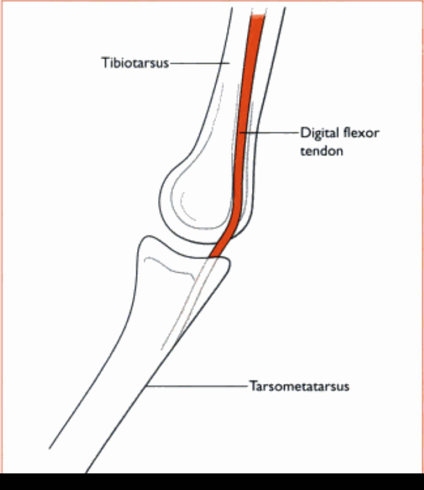
Fig. ∣3.4 Intertarsal joint ∙n the avian leg The digital flexor tendon runs in a tunnel within the metatarsal bone. It attaches to the caudal aspect of the digits and is important in the perching reflex.
wings of garden birds are good for slow' flapping flight: the w ings of barn owls have feathers with fringed edges w hich reduce the noise of the wingbeat as the owl approaches its prey.
Feathers
l eathers are the distinctive feature of members of the class Aves. They develop from epidermal cells in a similar way to the hairs of mammals and the scales of reptiles. Feathers arc made of keratin and create a strong but Iightwvight cowring over the w ing and the body.
All feathers have a similar structure (Hg. I 3.6). The central shaft or rachis is filled with blood capillaries during growrth but later, as the feather matures, it becomes hollow. On cither side of the shaft, the vane consists of barbs and interlocking barbules. These hook together to form a flattened wind-resistant surface.
The feathers must be kept dean in order to function effectively. Birds constantly groom themselves to zip up’ the barbules and to apply the secretions from the preen gland at the base of the tail, which keeps the feathers wraterproof.
'!‘here are four types of feather (Fig. 1 3.6):
1. Iliffht feathers - long rigid feathers attached to the wring and (he tail:
- Primaries: attached to digit 3 and to the fused metacarpal bones (Fig. 1 3.5). Usually 11 feathers but number varies with species: provide the major thrust during flight

F⅛. 13.5 Bird in flight showing the structure of the w∣ng (f?rel∣mb)
- Secondaries: attached to the ulna: shorter than the primaries
2. Contour feathers - cover the rest of the wing (coverts) and the outermost layer of the body to produce a smooth outline: shorter and more flexible: the lower part of the vane (closest to the skin) is fluffy.
3. Filopluineund
4. Down feathers - lie close to the body underneath the contour feathers, forming an insulating layer: they have no barbs so they are Huffy. Eiloplume is designed to break up. creating dust which absorbs sweat and dirt and keeps the bird clean.
Birds always shed dust from their feathers and this may be a source of pathogens. The zoonotic respiratory disease psittacosis is caused by the rickettsia Chlamydia psittaci and is spread by feather and faecal dust. Avoid inhaling dust from suspected cases.