Major skeletal muscles
There are over 600 skeletal muscles in the body. We will only cover the principal muscles, presenting them as groups controlling various parts of the body (see Table 7.4, Table 7.5, and Table 7.6 and Fig.
7.25, Fig. 7.26, Fig. 7.27, Fig. 7.28, and Fig. 7.29).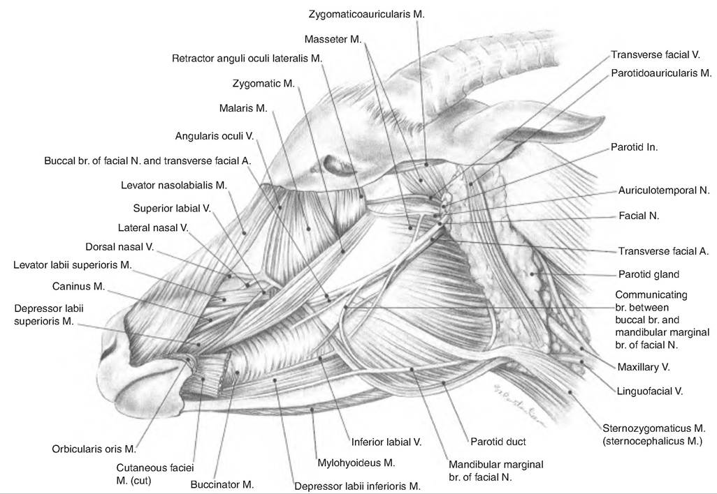
Fig. 7.25. Superficial muscles of the neck of the goat. A, artery; br, branch; V, vein; In, lymph node; M, muscle; N, nerve. (With permission from Constantinescu, 2001.)
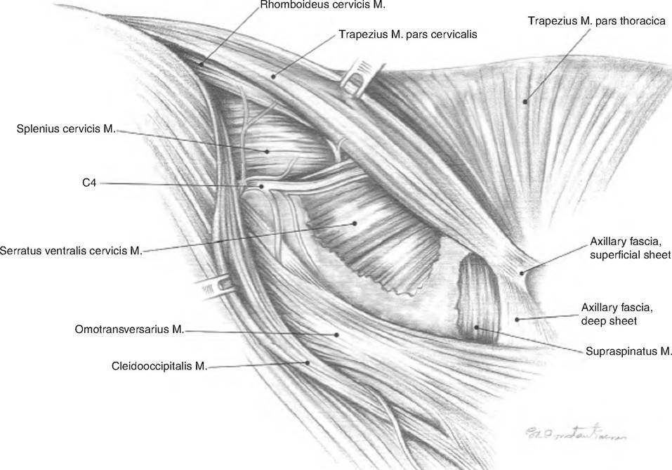
Fig. 7.26. Deeper muscles of the neck of the goat. A, arterty; V, vein; In, lymph node; M, muscle; N, nerve. (With permission from Constantinescu, 2001.)
Muscles of the pectoral limb and lateral thorax
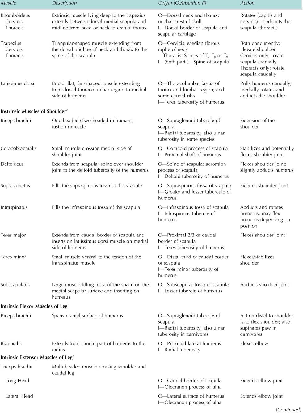
Table 7.5. Muscles of the pectoral limb.
| Muscle | Description | Origin (O)∕lnsertion (I) | Action |
| Extrinsic Muscles of Thoracic Limb1 | |||
| Brachiocephalicus | This is a wide muscle running from the head and neck to the front limb. The clavicular intersection divides the muscle into the Cleidocephalicus and Cleidobraehialis | Draws the lifted forelimb forward or draws the head to the side | |
| Cleidocephalicus | O—Occipital and dorsal midline of neck I—Clavicle | Pulls humerus forward | |
| Clavodeltoid | O—Clavicle I—Cranial border of humerus | Pulls humerus forward | |
| Pectoralis | A broad flat muscle extending from the sternum to the humerus | ||
| Pectoralis superficialis | O—Sternum I—Humerus (greater and lesser tubercles) | Adducts and retracts limb (flex shoulder) | |
| Pectoralis profundus | O—Sternum I—Cranial surface of the humerus | Flexes shoulder and draws humerus caudally | |
| Muscle | Description | Origin (O)∕lnsertion (I) | Action |
| Medial Head | O—Medial surface of humerus I—Olecranon process of ulna | Extends elbow joint | |
| Accessory Head | O—Proximal caudal humerus | Extends elbow joint | |
| (carnivores only) | I—Olecranon process of ulna | ||
| Tensor faciae antebrachii | Thin, insignificant muscle arising from the latissimus muscle | O—Latissimus dorsi I—Olecranon process of ulna and antebrachial fascia | Extends elbow joint |
| Anconeus | Small muscle filling olecranon fossa | O—Epicondyles of humerus | Lifts joint capsule out of |
| I—Olecranon process of ulna | joint space on full elbow extension |
1MuscIes that attach to the thoracic limb and some other part of the body.
2MuscIes having both attachments on the thoracic limb bones.
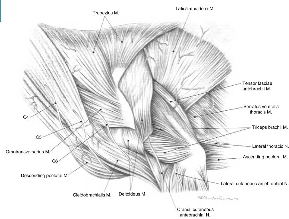
Fig. 7.27. Superficial muscles of lateral thorax of the goat. C, cervical spinal nerves; M, muscle; N, nerve. (With permission from Constantinescu, 2001.)
Muscles of the pelvic limb and body wall
Table 7.6. Muscles of the pelvic limb and body wall.
| Muscle | Description | Origin (O)∕lnsertion (I) | Action |
| Sublumbar Musdes1 | |||
| Psoas minor | A long, thin muscle extending from lumbar vertebral bodies to ilium | O—Vertebrae T13-L5 I—Iliopubic tubercle | Stabilize back; flex vertebral column |
| Iliopsoas (psoas major and iliacus) | Fused psoas major and iliacus muscle; chief flexor of hip | O—Lumbar vertebrae I—Lesser trochanter of femur | Flexes hip when leg is free; flexes vertebral column when leg is fixed |
| Quadratus Iumborum | Lies along underside of transverse processes of lumbar vertebrae | O—Transverse processes of lumbar vertebrae I—Wing of sacrum and ilium | Stabilizes lumbar vertebrae |
| Rump Musdes2 | |||
| Gluteal muscle | |||
| Superficial | Smallest, thinnest gluteal muscle | O—Sacral vertebrae I—Third trochanter | Abducts limb of femur |
| Medial | Largest gluteal muscle | O—Wing of ilium I—Greater trochanter of femur | Extends hip, abducts limb |
| Deep | Moderately sized, fan shaped | O—Body of ilium I—On or near greater trochanter | Extends hip, abducts limb |
| Tensor fasciae Iatae | Triangular-shaped | O—Tuber coxae I—Lateral femoral fascia, patella | Tense lateral femoral fascia and thus flex hop joint and extend stifle |
| Pelvic-Associated Muscles3 | |||
| Obturators | |||
| External | Fan-shaped muscle covering obturator foramen externally | O—Ventral surface of pubis and ischium surrounding obturator foramen I—Trochanteric fossa of femur | Adducts thigh |
| Internal | Fan-shaped muscle covering covering obturator foramen inside pelvic cavity | O—Interior (floor) of pelvis surrounding obturator foramen I—Trochanteric fossa of femur | Rotates femur laterally |
| Gemelli | Fairly insignificant hip rotator | O—Ischium I—Trochanteric fossa of femur | Rotates femur laterally |
| Quadratus femoris | Small rectangular muscle that extends from ileum to femur | O—Ventral surface of the caudal ischial tuberocity I—Intertrochanteric crest, just distal to intertrochanteric fossa | Outward rotation of the hip; minor action in extending hip |
| Body Wall Abdominal obliques | Four muscles forming ventrolateral abdominal wall | ||
| External | Sheet-Iike muscle extending Cranially and ventrally from ribs and thoracolumbar facia | O—Last several ribs and thoracolumbar fascia I—Midventral | Support the abdominal wall; assist forced expiration/ urination/defecation; twist trunk |
| Internal | Sheet-Iike muscle deep to the external abdominal oblique | O—Tuber coxae and thoracolumbar fascia I— Ventral midline at the Iinea alba | Support the abdominal wall; assist forced expiration/ urination/defecation; twist trunk |
| Gracilis | Broad, superficial muscle extending from pelvic symphysis to medial thigh and inserting on the tibia | O—Symphyseal tendon (ventral pelvic symphysis) I—Medial surface of knee/leg | Adducts thigh |
(Continued)
| Muscle | Description | Origin (O)∕lnsertion (I) | Action |
| Rectus abdominis Thigh Muscles4 | Tow long, straight muscles running from sternum to prepubic tendon | O—Sternum I—Prepubic tendon | SupportZbaIance |
| Hamstrings | |||
| Biceps femoris | Largest and most lateral of caudal thigh muscles | O—Ischial tuberocity I—Lateral surface of knee/leg | Extends thigh, flexes leg |
| Semimembranosus | Arises with semitendinosis muscle | O—Ischial tuberocity | Extends hip, flex or extend |
| form ischiatic tuberosity. Splits into two bellies. | I—Posterior surface of femur and tibia | knee (stifle) | |
| Semitendinosus | The longest hamstring; forms | O—Ischial tuberocity | Extends hip and tarsus, flexes |
| caudal border of thigh. | I—Tibia and calcanean tuberosity | knee (stifle) | |
| Pectinius | Long, spindle-shaped muscle on medial thigh | O—Pubic tendon I—Femoral shaft | Adducts limb and flexes hip |
| Quadriceps femoris | Large muscle covering lateral, medial and cranial surfaces of femur | Main stifle (knee) joint extensor | |
| Vastus lateralis | O—Cranial and inferior to greater trochanter of femur and along linea aspera I—Tibial tuberosity via patellar ligament | bgcolor=white>Extends knee (stifle) ||
| Vastus intermedius | O—Craniolateral surface of femur and linea aspera of femur I—Tibial tuberosity via patellar ligament | Extends knee (stifle) | |
| Vastus medialis | O—Entire length of linea aspera of femur I—Tibial tuberosity via patellar ligament | Extends knee (stifle) | |
| Rectus femoris | O—Rectus femoris area of ilium I—Tibial tuberosity via patellar ligament | Extends knee (stifle); flexes hip | |
| Sartorius | A strap-like muscle on medial thigh | O-Ilium I—Knee | Flexes hip joint and extends knee |
’Originates from ventral surface of caudal thoracic and lumbar vertebrae and inserts on the os coxae and femur.
2Originates from the ilium and inserts on the femur.
3Originiates Caudomedial to the hip joint and inserts in or near trochanteric fossa.
4IncIudes extensors of the stifle that are innervated by the femoral nerve, adductors and hamstring musles.
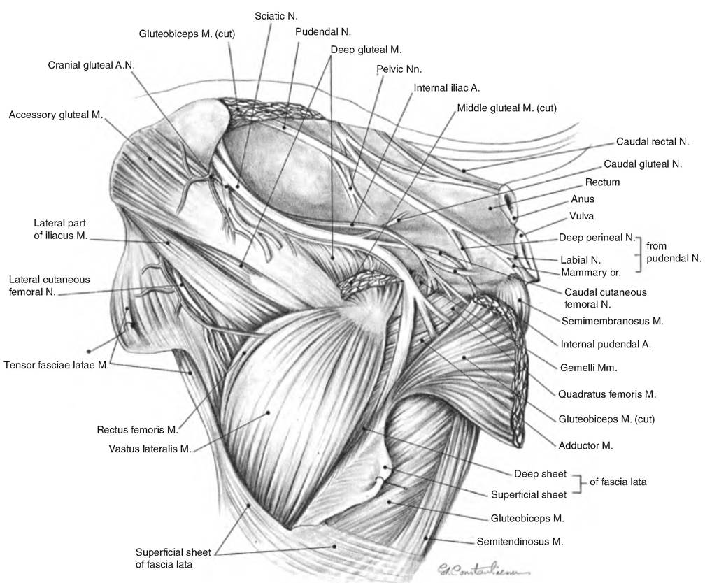
Fig. 7.28. Deeper muscles of left pelvis and thigh of goat, lateral aspect. A, artery; M, muscle; N, nerve; br., branch. (With permission from Constantinescu, 2001.)
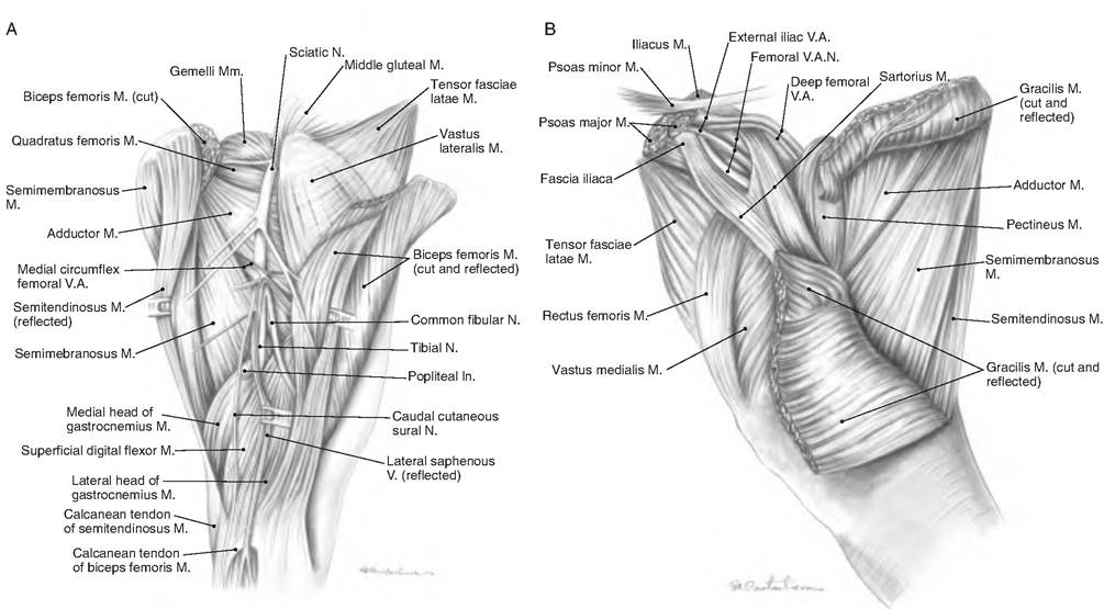
Fig. 7.29. Superficial and deep muscles in right thigh of goat. (A) Lateral aspect. (B) Medial aspect. M, muscle; N, nerve; V, vein. (With permission from Constantinescu, 2001.)
Stay apparatus of a horse
The stay apparatus allows a horse to rest while standing, using little muscular activity or fatigue. The stay apparatus uses a system of tendons and ligaments to "lock" the lower portion of the leg, thus requiring minimal muscular effort to stand (Fig. 7.30). The stay apparatus of the thoracic limb consists of the tendinous tissue of the serratus ventralis muscles, biceps brachii, lacertus fibrosus, radial carpal extensor, common digital extensor, long head of the triceps, suspensory ligament and its branches, collateral ligaments, superficial and deep digital flexor tendons, and their accessory ligaments. The stay apparatus works as follows:
1. Shoulder flexion. Prevented by the tendon of the biceps brachii.
a. Serratus ventralis. When the horse is at rest, the body is suspended from the scapulae by fibrous tissue in the serratus ventralis that causes the shoulder to flex.
b. Biceps brachii. The tendon of the biceps brachii runs the length of the muscle. Stretching this tendon prevents flexion of the shoulder.
2. Elbow flexion. The elbow tends to flex because of the weight of the animal.
This is prevented by placement of collateral ligaments behind the axis of the joint.3. Carpus flexion. The carpus would tend to flex, but this is prevented by the tendon of the biceps brachii, the lacertus fibrosus, and the tendon of the extensor carpi radialis acting cranially, and the flexor tendons and accessory ligaments acting Caudally
a. The lacertus fibrosus is a tendinous band that connects the tendon of the biceps brachii muscle to the tendon of the extensor carpi radialis muscle, thus forming an unbroken line of force from the shoulder to the metacarpus.
b. Tendon of the extensor carpi radialis. Tension is transmitted through the tendon of the biceps brachii and pulls the lacertus fibrosus, which directs tension down to the tendon of the extensor carpi radialis and then down to the metacarpus.
4. Carpus hyperextension. This is prevented by the cube shape of the carpal bones and the palmar carpal ligament.
5. Fetlock hyperextension. This is prevented by the suspensory apparatus, the extensor branches of the suspensory ligament, and the flexor tendons and their accessory ligaments.
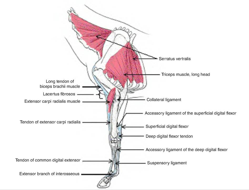
Fig. 7.30. Stay apparatus of the thoracic limb of the horse. (Modified from Pasquini et al., 1995.)