Muscle system
Naming muscles
The muscle system includes all the skeletal muscle and is therefore responsible for voluntary movement. It is not the intent of this book to cover all the muscles, but rather to give an overview of many of the major muscles involved in movement.
The muscles will be covered by functional area.Generally, the name of the muscle gives considerable information about its location or role. Muscles are named by several criteria:
1. Location. Sometimes, a bone or body region is included in the name, giving an indication of its location (e.g., temporalis is located over the temporal bone and the intercostal muscles are found between the ribs). The word deep (e.g., deep digital flexor) indicates that the muscle is not found at a superficial level.
2. Action. The action performed by the muscle may be included in the name. For example, the extensor digitorum extends a digit while the pronator teres pronates a limb.
3. Size. Whether the muscle is large or small may be indicated in the name (e.g., pectoralis major, adductor longus). The name may include such terms as Iongus (long), brevis (short), maximus (largest), minimus (smallest).
4. Shape. Sometimes muscles are named according to their shape (e.g., deltoid or trapezius muscles).
5. Direction of the fibers. Terms, such as rectus, indicate that the muscle fibers run parallel or straight relative to the body axis, whereas transverse muscles run at right angles to the same axis. Oblique fibers run at some other angle relative to that axis.
6. Number of origins/bellies. The name may include the number of heads (e.g., biceps brachii) or bellies (digastricus).
7. Attachment. Many times, the origin or attachment site is included in the name of the muscle (e.g., Cleidomastoid is attached to the clavical, or its remnant, and mastoid process).
Muscles can have several types of attachments.
They may attach to a bone via either (1) a tendon, which is a dense cord of regular connective tissue, (2) an aponeurosis, which is a tendinous sheet, or (3) fascia, which is common for superficial muscles, or (4) they may attach directly to the periosteum of the bone. Examples of each include the biceps brachii, which attaches via a tendon; the latissimus dorsi, which attaches via the thoracolumbar aponeurosis; the platysma, which attaches via the fascia; and the masseter, which attaches to the mandible.Arrangements of fascicles
As stated previously, skeletal muscles are arranged in fascicles (Fig. 7.1). The fascicle arrangement can vary (Fig. 7.21). Fascicles are sometimes found to be arranged in a straight line and are referred to as parallel muscles (an example is the abductor digiti minimi). If the arrangement is circular, the fascicles appear as concentric rings. Such an arrangement is typically
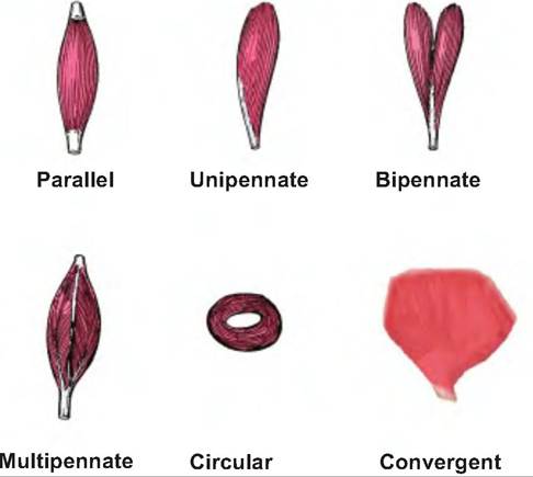
Fig. 7.21. Fascicle arrangement. Muscle fascicles can have varying arrangements allowing for different functions.
found around an orifice such as the orbicularis oris muscle that surrounds the mouth. In a convergent muscle, the fascicles converge to the tendon for insertion. Therefore, the muscle begins wide, similar to the shape of a fan, and then narrows, as is seen in the pectoralis major muscle. In the pennate muscle, such as the gastrocnemius muscle, the fascicles are short and attach obliquely (penna = feather) to the tendon. In a unipennate muscle, the tendon runs along one side of the tendon, whereas in multipennate, the tendon branches within the muscle and looks like many feathers laying side by side.
Muscles as levers
The muscular and skeletal systems work together to function as a lever system. A lever consists of a rigid structure (i.e., bone) that moves around a fixed point called the fulcrum. Muscle contraction acts as the force to move a load, or bone.
Levers provide a mechanical advantage, allowing a force to move a heavier load either further or faster (Fig. 7.22).Three different classes of levers exist, depending on the relationship of the force, fulcrum, and load (Fig. 7.23). In a first-class lever, the fulcrum is between the force and load, which are at either end of the lever. This is similar to a playground seesaw, and it provides a mechanical advantage. The act of an animal lifting its head functions as a first-class lever.
A second-class lever exists when the force is applied at one end and the fulcrum is found at the other end, while the load is placed in between. Such levers create a mechanical disadvantage, and are thus rarely found
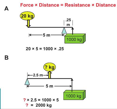
Fig. 7.22. Mechanical advantage versus disadvantage. A lever system can provide a mechanical advantage or a mechanical disadvantage. The equation shown at the top explains the relationship between force and distance. (A) With this lever system, a mechanical advantage is provided because of the position of the fulcrum relative to the force and load. Twenty kilograms of force is able to lift 1OOOkg of load. (B) With the fulcrum and load placed at opposite ends of the lever, and a force provided in the middle, it requires 100 times the amount of force needed in the example shown in A to move the same load. This is a mechanical disadvantage.
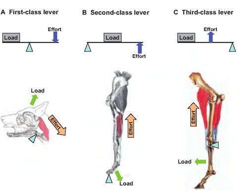
Fig. 7.23. Anatomical examples of lever systems. (A) In a first-class lever, the force and load are on opposite sides of the fulcrum. (B) In a second-class lever, the load is between the force and the fulcrum. (C) In a third-class lever, the force is between the load and the fulcrum.
in the body. A wheelbarrow acts as such a lever. The calf muscles act as a second-class lever when causing plantar flexion.
In a third-class lever, the fulcrum and load are at either end of the lever, and the force is applied between the two.
Tweezers work in this manner, as do most skeletal muscles. When the biceps brachii flexes the elbow, the insertion is located on the radius-ulna while the origin is located in the shoulder region. Contraction of the muscle causes the leg to flex at the knee. In a third-class lever, the speed and distance traveled by the load are increased at the expense of effective force.Muscle terminology
Origins and insertions
Muscles are always attached to other structures at either end. Generally, one end is in a fixed position while the other end moves toward it when the muscle is contracted. The fixed attachment site is called the origin, and the movable end is called the insertion (Fig. 7.24). While the origin is normally proximal or superior to the insertion, this is not always the case. The sternocleidomastoid muscle originates at the sternum, and inserts at the mastoid process of the temporal bone.
Actions
The movements of the skeleton caused by muscle contraction involve flexion or extension, adduction or abduction, protraction or retraction, elevation or depression, rotation, circumduction, pronation or supination, inversion or eversion. These actions are defined in Table 7.3.
Muscles typically work in groups rather than individually. For example, the biceps branchii and brachialis cause flexion of the elbow, and the triceps brachii, tensor fasciae anterbrachii, and anconeus cause extension of the elbow. The agonist is the muscle primarily responsible for producing a certain movement, and the antagonist is the muscle whose action opposes that movement. In the case of the elbow, the
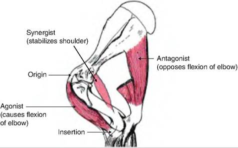
Fig. 7.24. Muscle functions and attachments. Using the thoracic limb of the horse, this figure demonstrates the origin and insertion of muscles, as well as the agonist and antagonist functions with regards to the elbow. (Figure modified from Getty, 1964.)
| Term | Description |
| Abduction | Movement of a structure or limb or away from the median plane of the body (e.g., spreading the toes or moving a limb away from the center of the body) |
| Adduction | Movement of a structure or limb toward the median plane of the body (e.g., bringing the toes together or moving a limb toward the center of the body) |
| Antagonist | Opposes the movement of the prime mover (e.g., the triceps brachii, which extends the elbow joint, is the antagonist of the biceps brachii, which flexes the elbow joint) |
| Circumduction | Circular movement of an extremity describing the surface of a cone; involves successive flexion, abduction, extension, and adduction (e.g., moving a limb in a circular motion) |
| Depression | Moving a structure in a ventral direction (e.g., dropping the shoulders) |
| Elevation | Moving a structure in a dorsal direction (e.g., shrugging the shoulders upward) |
| Eversion | Twisting the foot to face the sole outward |
| Extension | Increasing the angle between bones. Ex: extending fingers from a curled position, or straightening the stifle (knee) |
| Flexion | Decreasing the angle between bones. Ex: Bending the elbow toward a 90o angle. |
| Trunk lateral flexion | Bending to the side |
| Trunk flexion | Bending forward |
| Shoulder flexion | Swinging the limb backward (in humans, it is swinging the limb forward) |
| Plantar flexion | Movement of the ankle joint to push the sole of the foot downward |
| Dorsiflexion | Flexion of the ankle joint in order to raise the top of the foot upward |
| Insertion | The more movable of the two attachments. In the limbs, this is usually the more distal attachment |
| Inversion | Twisting motion of the foot to face the sole inward |
| Origin | The less movable of the two attachments. In the limbs, it is usually the more proximal attachment |
| Pronation | Movement of the palmar side of the paw downward |
| Protraction | Moving a part of the body Cranially in the horizontal plane. Ex: Pushing the shoulders forward |
| Retraction | Moving a part of the body caudally. Ex: Pulling the shoulders back |
| Rotation | Movement around the long axis |
| Lateral rotation | The cranial surface of a limb turns away from the long axis of the trunk (e.g., Gemelli) |
| Medial rotation | The cranial surface of a limb turns toward the long axis of the trunk |
| Supination | Movement of the forearm (radius and ulna) so the palmar side is rotated upward or forward as when a cat laps milk off its paw. |
| Synergist | A muscle that indirectly aids the action of the prime mover. Ex. Teres major muscle is a synergist to the latissimus dorsi muscle |
Muscles of the head and neck
Table 7.4. Muscles of the head and neck.
| Muscle | Description | Origin (O)∕lnsertion (I) | Action |
| Muscles of Facial Expression and Mastication | |||
| Buccinator | Muscle contained within the wall of | O—Fascia of cheek wall | Holds the cheeks toward the |
| the cheek | I—Fascia of cheek wall | mouth when chewing | |
| Masseter | Most powerful muscle for closing jaw | O—Zygomatic arch | Elevates mandible (closes |
| I—Masseteric fossa of mandible | mouth) | ||
| Mentalis | Major muscle making bulk of muscle | O—Rostral end of mandible | Elevates and protrudes lower lip |
| mass of chin | I—Skin of chin | ||
| Orbicularis oris | Multilayered muscle of the lips | O—Lips I—Lips | Purses lips |
| Pterygoid | |||
| Medial | Two-headed muscle running along | O—Sphenoid and pterygoid bones | Acts with temporalis and |
| internal surface of mandible | I—Medial surface of mandible | masseter muscle to elevate mandible (close mouth) | |
| Lateral | Two-headed muscle lying superior to | O—Sphenoid bone | Protrudes mandible; in |
| medial pterygoid muscle | I—Neck of condyle of mandible | herbivores, cross jaw-action | |
| Platysma | "Grimace" and "neck-cording" muscle | O—Skin of neck I—Angle of lips | Retract lips |
| Temporalis | Fan-shaped muscle covering part of | O—Temporal fossa | Closes mouth; elevates and |
| temporal, frontal and parietal bones | I—Coronoid processs of mandible | retracts mandible | |
| Zygomaticus | Muscle running diagonally from cheek bones to corner of mouth | O—Zygomatic bone near zygomaticomaxillary suture I—Angle of the mouth | Retracts and elevates corners of mouth |
| Muscles of the Neck | |||
| Brachiocephalicus | |||
| Cleidobrachialis | Complex muscle acting on head and/ | O—Clavicle or its remnant | Depending on which end is |
| or limb | I—Cranial humerus | fixed (not moved), and whether | |
| Cleidomastoideus | O—Clavicle or its remnant I—Mastoid process of skull | one or both sides (right and left) contract: Advances forelimb, | |
| Cleidocephalicus | O—Clavicle or its remnant I—Dorsal midline of neck | turns head to the side, or flexes neck ventrally | |
| Sternocephalicus | In carnivores, consists of | O—Cranial part of the sternum | Flexes the head and neck and |
| Sternomastoid and Sternoocipitalis. In | (manubrium) | inclines them to one side. | |
| ox and goats, the Sternoocipitalis is | Sternomandibularis may also | ||
| replaced by the Sternomandibularis. The horse has only Sternomandibularis. | I—Mastoid process of skull | help open the mouth. | |
| Sternomastoid | O—Sternum (manubrium) I—Mastoid process of skull | ||
| Sternooccipitalis (dog and cat) Sternomandibularis | O—Sternum (manubrium) I—Occipital region of skull O—Sternum (manubrium) I—Angle of the mandible | ||
biceps brachii is the agonist for flexion of the elbow and the triceps branchii is the antagonist. A synergist is a muscle that helps the agonist work more efficiently. Synergists may provide additional force to move the joint, or they may help stabilize the joint.
Muscle attachments
Muscles can make three types of attachments. In a fleshy attachment, there is an apparent direct attachment of the muscle to the bone, as in the case of muscle attachments to the scapula. In actuality, the muscles are attached to the bones by very short tendons. In a
tendinous attachment, the muscle is attached to the bone via dense connective tissue. In an aponeurotic attachment, there is a flat, tendinous sheet attaching the muscle, as is seen in the abdominal wall.
More on the topic Muscle system:
- TECHNICAL FACTORS OF NEEDLE ELECTROMYOGRAPHY
- 31 Urogynecology and Reconstructive Pelvic Surgery
- Dystrophic Myopathies
- Agrawal M.. Textbook of Pediatrics. 3rd ed. — CBS Publishers,2025. — 973 p., 2025
- Inspection
- System Models of Communication
- Neoplasia of the Reproductive System
- Skeletal System
- Pain control
- Respiratory Failure