Smooth muscle
Smooth muscle, also called nonstriated or involuntary muscle, can be found surrounding the blood vessels, digestive tract, urinary system, reproductive system, and respiratory system.
It can be found in the form of bundles or sheets around other tissues.Structure
Smooth muscle cells are long and slender, varying from 5 to 10 μm in diameter and 30-200 μm in length, with a single, centrally located nucleus. There are no T tubules, and the sarcoplasmic reticulum is not as well organized as in skeletal and cardiac muscle. As discussed in the following, the source of Ca2+ for smooth muscle contraction is mostly the extracellular space. To facilitate the entry of Ca2+, the Sarcolemma of smooth muscle has in-foldings called caveoli that increase the surface area.
Smooth muscle also lacks the well-organized connective tissue sheaths found in skeletal muscle. There is an endomysium found between smooth muscle cells that is secreted by the smooth muscle cells and that contains blood vessels and nerves.
While smooth muscle contains actin and myosin, it lacks myofibrils and sarcomeres that cardiac and skeletal muscles possess. As a result, smooth muscle lacks striations, hence the name smooth or nonstriated muscle.
Thick filaments are found throughout the smooth muscle cell. Cross bridges are more numerous in smooth muscle since they are found along the entire length of the myosin filament, which is also longer than in skeletal muscle. In addition, scattered throughout the sarcoplasm is a network of intermediate filaments composed of the protein desmin (Fig. 7.19). Attached to the intermediate fibers are structures called dense bodies. Some dense bodies attach directly to the Sarcolemma, and the thin filaments are attached to dense bodies. Therefore, dense bodies act similarly to the Z discs in striated muscle.
The ratio of thin to thick filaments is much lower in smooth muscle (1:13) than in skeletal muscle (1:2). However, the thick and thin filaments do interdigitate.Due to the network of attachments between the thin filaments and the sarcolemma, when the thick filaments pull on the thin filaments, this causes the cell to shorten, with the areas between the dense bodies bulging outward, creating an irregular cell surface (Fig. 7.19). Since the network of intermediate fibers crosses throughout the cell, during contraction the cell does not simply shorten in one plane, but rather shortens in multiple directions.
Types of smooth muscle
There are two types of smooth muscle: single-unit smooth muscle and multiunit smooth muscle (Fig. 7.20).
1. Single-unit smooth muscle. Single-unit smooth muscle, also called visceral smooth muscle, is widely distributed throughout the body. The cells are electrically coupled by gap junctions, allowing an electrical impulse to move between cells. Since the cells are electrically connected, they function
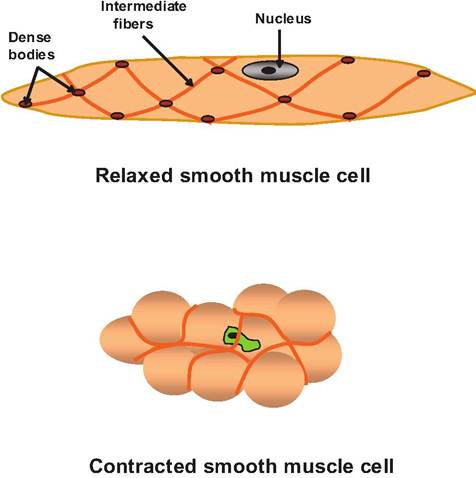
Fig. 7.19. Smooth muscle cell. A smooth muscle cell is long and slender, with a single nucleus. Unlike the skeletal muscle fibers, the smooth muscle fibers lack cross striations. However, smooth muscle fibers have intermediate fibers attached to dense bodies. Many of the dense bodies are attached directly to the sarcolemma. Since the intermediate fibers crisscross the cell, when stimulated, a smooth muscle fiber contracts in multiple directions.
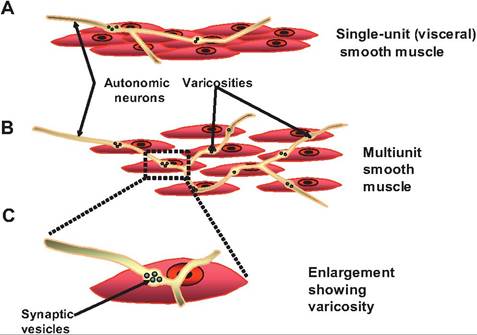
Fig. 7.20. Types of smooth muscle, and their innervations. (A) In single-unit, or visceral, smooth muscle, there are gap junctions between the individual muscle cells. Therefore, when one cell is stimulated by an autonomic neuron, the action potential can spread among cells so that single-unit smooth muscle can contract as a unit.
(B) In multiunit smooth muscle, individual muscle cells are separated from one another; therefore, each fiber needs its own innervation. (C) The autonomic fibers that innervate smooth muscle have varicosities along their length that make diffuse synaptic connections with muscle cells releasing neurotransmitters at these locations.as a unit in which an entire sheet of cells are interconnected. Such muscle is found along the wall of the digestive tract, the gall bladder, the urinary bladder, and most other internal organs.
This type of muscle is often found as two layers: a longitudinal layer running parallel to the long- axis of the organ and a circular layer in which the fibers encircle the organ. When the longitudinal layer contracts, this dilates and shortens the organ, whereas contraction of the circular layer constricts the lumen of the organ. Such muscle generally displays rhythmic contractions that are controlled by pacesetter cells that can spontaneously depolarize and trigger contraction of the remainder of the muscle.
2. Multiunit smooth muscle. In contrast to singleunit smooth muscle, individual cells are separated from one another in multiunit smooth muscle. Such cells generally lack gap junctions. This necessitates that each cell must be innervated by a nerve ending. Therefore, such muscle has a richer nerve supply than single-unit smooth muscle. There is seldom synchronous contraction of the entire muscle. This type of muscle is found in the iris of the eye, along portions of the male reproductive tract, surrounding the walls of large arteries, and in the arrector pili muscle of the skin.
Neural innervation of smooth muscle
Smooth muscle is innervated by the autonomic nervous system, whereas skeletal muscle is innervated by the somatic nervous system. Unlike skeletal muscle that has a well-defined NMJ, autonomic fibers run over the surface of smooth muscle cells and have many bulbous swellings called varicosities. The neurotransmitter is released from these varicosities and diffuses into a wide synaptic cleft, forming a diffuse junction.
Contraction of smooth muscle
Contraction of smooth muscle takes longer to develop, but lasts longer, than skeletal muscle. Smooth muscle can also shorten and stretch further than skeletal muscle. This is due to the differences in the structure between the two muscle types. While tropomyosin is found in smooth muscle associated with the thin filaments, troponin is lacking. There is less SR in smooth muscle, and smooth muscle lacks T tubules. As a result, the action potential must spread only along the surface of the cell, and the greatest source of calcium for muscle contraction is the interstitial space. The sequence of events for smooth muscle contraction is as follows:
| Table 7.2. Comparison of skeletal, cardiac, and smooth muscle. | |||
| Properties | Skeletal Muscle | Cardiac Muscle | Smooth Muscle |
| Location | Attached to bones; or occasionally to skin in the case of some facial muscle | Wall of the heart | Single-unit found in walls of hollow organs; multi-unit found in intrinsic ocular muscles |
| Size | Single, long, cylindrical; Striated; 100μm-30cm | Branching; Striated; 10-20μm ? 50-100μm | Fusiform; non-striated; 5-10μm ? 3O-2OOμm |
| Number of nuclei | Multinucleated | Uni- or binucleated | Uninucleated |
| Connective tissue components | Epimysium, perimysium, and endomysium | Endomysium attached to fibrous skeleton | Endomysium |
| Presence of sarcomere | Yes | Yes | No; actin and myosin scattered throughout sarcoplasm; actin attached to dense bodies |
| Presence and location of T tubules | Yes; located at junction of A and I bands | Yes; located at Z disc | No; caveoli (infolding of cell membrane) |
| Presence gap junctions | No | Yes at intercalated discs | No; caveoli instead |
| Individual neuromuscular junctions | Yes | No | Present in multiunit but not single-unit muscle |
| Source of calcium | Sarcoplasmic reticulum | Sarcoplasmic reticulum and extracelluar | Mostly extracellular |
| Site of calcium binding and regulation | Troponin on thin filaments | Troponin on thin filaments | Calmodulin in sarcoplasm |
| Contraction | Rapid onset, can tetanize depending on fiber type can fatigue rapidly | Develops slowly cannot tetanize | Slow onset, may tetanize but generally fatigues slowly |
| Effect of nervous system | Yes; located at junction of A and I bands | Yes; located at Z disc | No; caveoli instead |
| Respiration | Aerobic and anaerobic | Aerobic | Primarily aerobic |
1.
The muscle is stimulated by autonomic fibers releasing either norepinephrine or ACh.2. An action potential spreads along the Sarcolemma.
3. Cytosolic Ca2+ levels increase, with calcium coming mostly from the interstitial space, but some from the SR.
4. Ca2+ binds to the calmodulin, a second messenger, causing its activation.
5. The activated calmodulin then activates myosin light-chain kinase.
6. The activated protein kinase phosphorylates the myosin head.
7. Phosphorylation allows cross bridges to form between myosin and actin.
8. The muscle contracts as Ca2+ is pumped out of the cell.
Note that in smooth muscle, calmodulin is serving a role similar to that of troponin in skeletal muscle in that it binds calcium. Once activated, calmodulin activates a myosin light-chain kinase that uses ATP to phosphorylate the myosin head, thus allowing the myosin head to attach to actin. The pumping of calcium out of the cell is a slow process partially explaining the delay in smooth muscle relaxation.
In addition to responding to the autonomic nervous system, smooth muscle can contract and relax in response to stretching; hormones; or changes in local factors, including pH, O2, CO2, temperature, and ion concentrations. For example, stretching of the bladder can cause contractions. Hence, the bladder can continue to function in an animal with a spinal cord injury. Epinephrine, released from the adrenal medulla during a stress response, can cause the bronchioles to dilate. For a comparison of skeletal, smooth, and cardiac muscle (discussed in Chapter 13), see Table 7.2.