Mammary Fibroadenoma
Mammary fibroadenomas are a common occurrence in older female rats, particularly SD rats, although these tumors may also occur occasionally in male rats. Sex, age, genetic, dietary, and endocrine factors may all play a role in the incidence of spontaneous mammary tumors.
There are significant variations in the incidence, depending on the source of rats of this strain. Ranges of 7-40% have been recorded in SD rats from different sources. The incidence of spontaneous mammary fibroadenomas is markedly reduced in ovarectomized rats. Restriction of food intake by 20% reduced the incidence of mammary tumors in female SD rats by fivefold, compared with controls. Prolactin levels have also been identified as a key factor. Serum prolactin values in females with mammary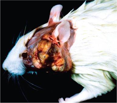
FIG. 2.76. Lobulated tumor arising from the Zymbal's gland. The Zymbal's gland is located within the subcutis adjacent to the ear, with ductal drainage into the external ear canal.
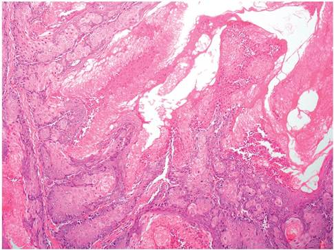
FIG. 2.77. Zymbal's gland tumor composed of polyhedral cells with central keratinization and cellular debris.
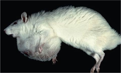
FIG. 2.80. Mammary fibroadenoma in an aged rat. These tumors will often grow to very large size without malignant transformation or metastasis.
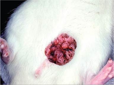
FIG. 2.78. Preputial gland adenoma in a mature male rat. Note the distinct lobulated appearance to the tumor. (Source: T.R. Schoeb, University of Alabama, Tuscaloosa, Alabama. Reproduced with permission from T.R. Schoeb.)
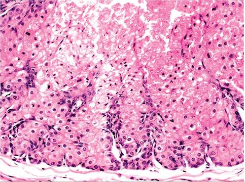
FIG.
2.79. Preputial gland tumor shown in the previous figure. The mass consists of well-differentiated glandular epithelial cells. (Source: T.R. Schoeb, University of Alabama, Tuscaloosa, Alabama. Reproduced with permission from T.R. Schoeb.)tumors were over 25 times higher than in 6-month-old virgin females. There have been attempts to equate mammary tumors with the incidence of pituitary adenomas, but unequivocal correlations have not been made.
Mammary fibroadenomas may reach very large size (Fig. 2.80), and are typically circumscribed, movable, firm, and lobulated. They may be located within any of the 12 mammary glands along the mammary chain, and occasionally at other sites on the body. In larger tumors, there may be ulceration of the overlying skin. On cut section, tumors are lobulated (Fig. 2.81), with regions of highly fibrous to glandular tissue. The lobu- lated appearance is readily evident on cut surface. On microscopic examination, there is distinct interlobular and intralobular connective tissue surrounding relatively well-differentiated acinar structures (Figs. 2.82 and 2.83). There are markedly variable proportions of acinar and collagenous tissue, depending upon region of the tumor examined. Acini are lined by cuboidal epithelial cells, frequently with prominent vacuoles in the cytoplasm.
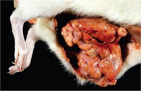
FIG. 2.81. Mammary fibroadenoma in an adult female Sprague-
Dawley rat. The prominent lobulations and interlobular fibrous tissue are characteristic gross findings seen with this neoplasm.
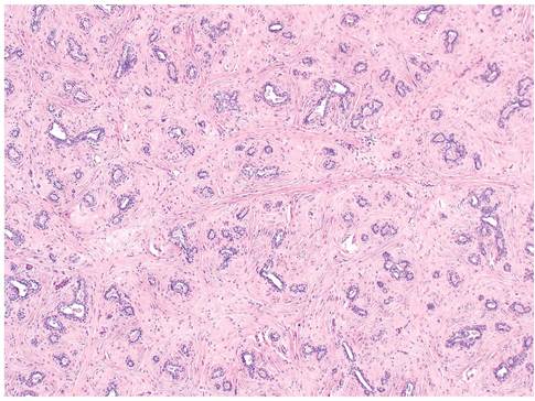
FIG. 2.82. Mammary fibroadenoma, illustrating the acinar structures and prominent connective tissue components.