Myocardial Degeneration/Fibrosis
Focal to diffuse areas of myocardial degeneration and fibrosis are frequently seen microscopically in
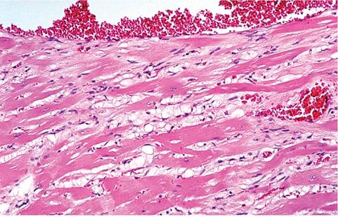
Fig.
2.55. Myocardial degeneration and interstitial fibrosis in an aged rat.conventional and specific pathogen-free rats, particularly after 1 year of age. Lesions are more common in male rats. The prevalence may be over 80% in some rat strains. At necropsy, there may be moderate to marked ventricular hypertrophy, and pale streaks may be evident on the epicardium. On microscopic examination, degenerative changes are usually most evident in the papillary muscles of the left ventricle, although the interventricular septum may also be involved. Atrophy of myofibers, vacuolation and fragmentation of sarcoplasm, loss of cross-striations, mononuclear cell infiltration, and fibrosis are typical changes (Fig. 2.55). Large reactive nuclei are occasionally observed. Interstitial fibrosis is an important feature of the disease, which is particularly evident in Trichrome-stained tissue sections (Fig. 2.56). Although this condition is frequently present in older rats, there may be little or no evidence of cardiac insufficiency.
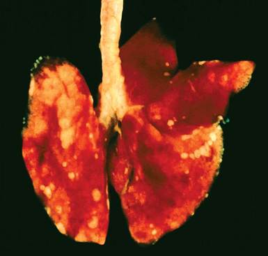
FIG. 2.54. Alveolar histiocytosis in an aged rat. Note the raised pale foci in the subpleural regions consistent with intra-alveolar aggregations of macrophages.
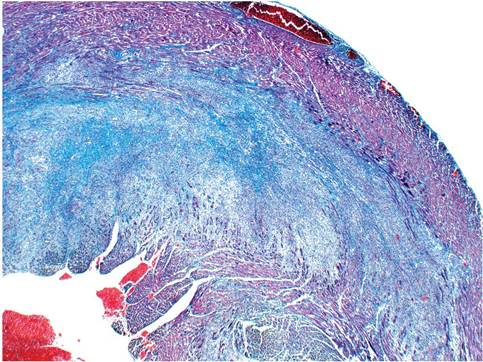
FIG. 2.56. Left ventricle of an aged rat with marked myocardial fibrosis. Note the abundant blue-stained collagen. (Masson's trichrome stain).
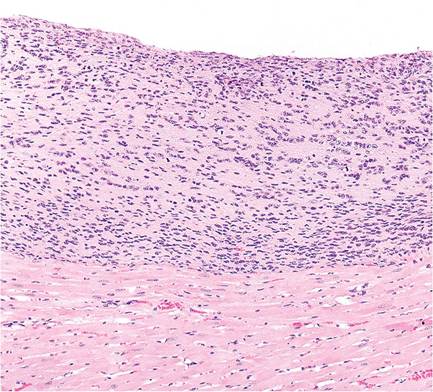
FIG. 2.57. Endocardial spindle cell proliferation lining the ventricular lumen (top). This spontaneous lesion may arise in various strains and stocks of rats.