Nails, claws, hoofs, and feathers
Let us begin by first considering the nails of humans and other primates. Nails are hard plates of tightly packed keratinized cells. They are clear and cover the dorsal surface of the last phalanges of fingers and toes.
Each nail is made up of three regions: (1) the nail body, (2) free edge, and (3) the nail root. The body of the nail is the visible portion. It rests on the nail bed, essentiallyBox 5.3 Rickets, anyone?
Rickets, considered a classic metabolic disease of bone, was first described in both humans and animals more than 2000 years ago. The discovery that vitamin D could prevent the disease led to a dramatic decrease in developed countries, but cases still occur. In humans, reduced sun exposure and increased use of sunscreen are believed to play a role. Dittmer and Thompson (2011) describe animal models that have been developed to study vitamin D physiology and related bone disease as well as etiology of rickets in domestic animals. This is most often associated with phosphorous deficiency or other genetic disturbances.
Skin, of course, is a primary physical barrier to protect the body from a variety of environmental pathogens, but specific secretions are also important. Fukui et al. (2012) report on the expression of a variety of naturally occurring antimicrobials, including lysozyme, defensin-2, lactoferrin, and others within the cells, and secretions of eccrine glands of the skin of the porcine snout. It is not hard, given the propensity of pigs to root and explore their environment, to imagine the importance of these products.
a combination of the stratum basale and the stratum spinosum. The free edge is that part that extends past the end of the digit. The nail root is hidden from view and is embedded in a fold of skin (or nail fold). The cuticle is the stratum corneum of the nail fold that gets pushed outward over the surface of the nail body.
Nail growth depends on the nail matrix located under the nail root. As you might guess from your review of the epidermis, the nail matrix consists of the two deepest layers of the epidermis (stratum basale and stratum spinosum). The keratinization of the cells of the nail matrix develops directly from the spinosum so that the stratum granulosum and Iucidum are absent. The result is the creation of a hard, durable plate. As the nail matrix proliferates and the cells are keratinized, this hard plate gets pushed forward onto the nail bed and the nail lengthens. If you consider your own nails, you can see a small white crescent. This is called the lunula, and it corresponds with the presence of the thick matrix underneath. So how does this differ from claws and hoofs?
We will use the horse as an example of the remarkable development of hooves. Let us begin with the lower leg and foot. The foot includes the hoof (epidermis) and the underlying dermis and associated structures. Specifically, these include the corium or dermis, digital cushion, terminal phalanx or coffin bone, the distal end of the second phalanx (short pastern bone), navicular bone, and a variety of muscles, tendons,
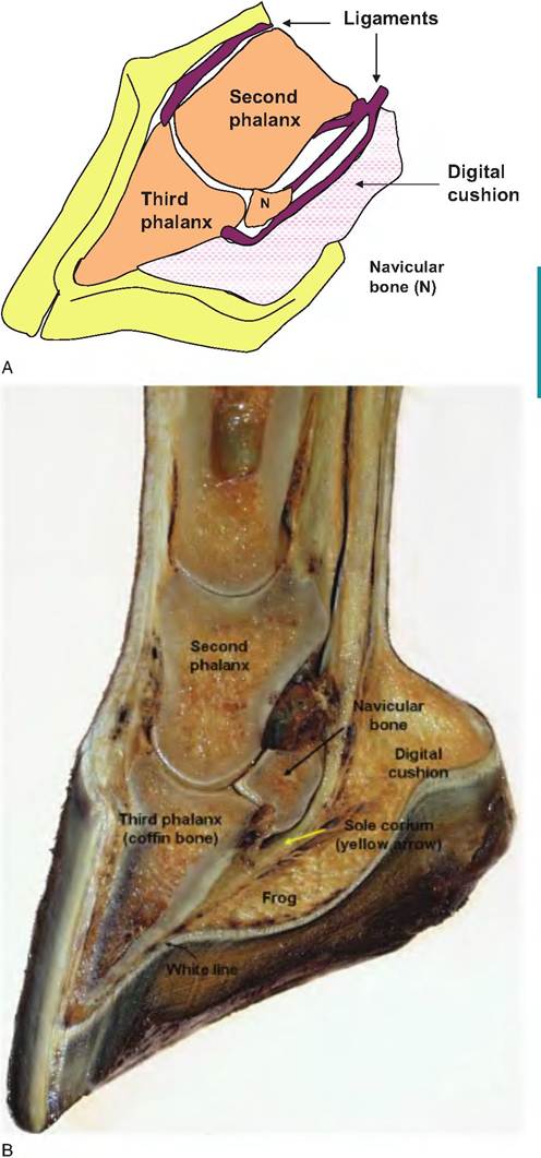
Fig. 5.9. Equine foot and hoof. Diagram (A) and corresponding photograph of cross section (B) of the equine foot.
ligaments, and nerves. Some of these major features are illustrated in Figure 5.9.
The hoof is largely an insensitive cornified layer derived from the epidermis. Strictly speaking, the terms "epidermis" and "dermis" are more accurate than the common terms "insensitive" and "sensitive," respectively. The structure of the hoof is produced in two fashions. In some regions, the dermis is very highly papillated; this mirrors the general appearance
of dermal papillae in other regions of the body, but they are more abundant and highly interlinked with corresponding epidermal pegs.
This organization increases strength and assists interchange between the dermal blood vessels and the avascular epidermis. In other areas, the papillae and epidermal pegs are so confluent that the adjacent epidermal and dermal tissues create distinct layers or lamina.The hoof is essentially an extension of the skin of the lower limbs. This boundary area that circles the leg just above the hoof is called the coronet. In this region, the dermis of the skin is continuous with the dermis or corium of the hoof. The subdivisions of the corium correspond with the adjacent epidermal regions of the hoof. As you move from the anterior surface of the hoof in a caudal direction, you pass through the following epidermal layers: stratum tec
torium, stratum medium, and stratum internum. The stratum tectorium extends from the periople layer of the skin epidermis in the area of the coronet. Most of the wall is made up of the stratum medium and is organized in a tubular arrangement of cells. This is similar in appearance to compact bone if cut in cross section. These tubular structures are sometimes noticeable as lines oriented from the coronet toward the ground in the wall of the hoof. The boundary between the outer "insensitive" epidermis and the "sensitive" dermis occurs in the stratum internum. Here there is a complex interaction between the outer epidermis and underlying laminar corium. Specifically, papillae from the dermis extend into the epidermis in a regular repeating array. Beyond the dermis (depending on the angle) you would penetrate the bone of the distal phalanx. This is illustrated in Figure 5.10. If you penetrated from the bottom of the hoof near the center, you would pass through similar (but thinner) layers of epidermis and the tubular and intertubular horn of the sole, the solar corium (dermis), the periosteum, and compact bone of the distal phalanx.
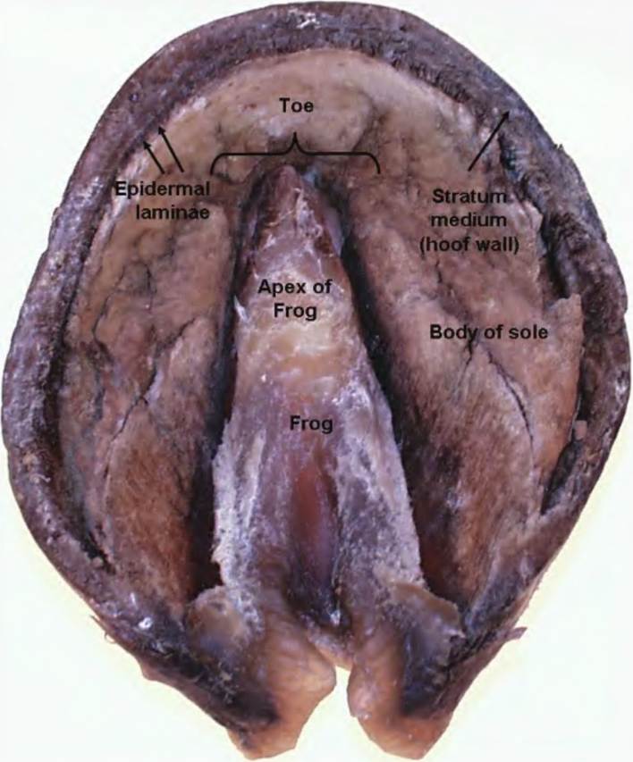
Fig.
5.10. View of the plantar (bottom) of the equine hoof.In addition to these complex histological structures, there are also a number of well-defined structures associated with the hoof. These include the frog (so named because of its shape), bulbs, and the sole. The anterior edge is the toe and the caudal margin the heel. The white line, most apparent when the hoof is trimmed, is created by the epidermal laminae and represents the boundary between the dermis and epidermis. The frog is a wedge-shaped mass of mostly keratinized tissue that is softer than in other areas because of higher water content. The tubules of the sole are arranged vertically in correspondence with the papillae of the dermis or corium of the sole. The corium itself has a good supply of nerve and blood vessels. Figure 5.10 illustrates the gross anatomy of an equine hoof. The photograph is of the bottom or ventral surface of a freeze-dried hoof. It is easy to imagine the structure like a shoe that surrounds the dermis bones, blood vessels, and nerves of the lower foot.
Horns
The horns of cattle, goats, and sheep are generated in the region over the horn process, a germinal center that projects from the surface of the frontal bone of the skull. The dermis or corium envelops the horn core so that it is fused with the outer covering of the bone tissue or periosteum. As the horn develops, the corium at its base is especially thick where it links to the skin via abundant long slender papillae. The papillae become shorter and less abundant toward the apex of the horn. The bulk of the horn tissue consists of tubules that extend from the base of the horn toward the apex. Softer tissue covers the surface of the horn near the base and extends a variable distance toward the apex. This is similar to the outer covering of the hoof, the periople. Growth of the horn varies with nutrition. This can be especially evident in wild animals with seasonal variation in available nutrients. This variation is reflected in the appearance of rings in the horn that can be used to estimate age.
In many commercial situations, especially dairy, polled cattle are often dehorned. This is usually accomplished by destroying the corium when only the horn buttons are present. This is typically accomplished by either surgical removal or destruction with a hot iron or application of caustic paste. Once the horn has begun to develop, both the corium and entire horn must be removed to prevent the horn from redeveloping. Even a small amount of corium can produce a crooked, stubbed horn.
Feathers
Feathers are likely the most complex structure that is derived from the integument among vertebrates. It is also clear that there are dramatic differences between avian species as well as marked differences depending on location and purpose. To illustrate, the tail feathers of a rooster are more than 1000 times larger than the smallest feathers on his body. First let us consider what feathers actually do. Protection from the elements and maintenance of body temperature are clearly critical in all birds, but in addition, they allow the bird to fly. Other aspects include protection from predators and the ability to attract the members of the opposite sex. Feathers are also plentiful. A mature chicken has about 5000 feathers.
Feathers come in several forms, but they are all made up of the same basic parts; however, these parts may be absent or rearranged from type to type. The predominant feather type on a bird's body is called the pennae or contour feather. Other feathers vary from this common type to large stiff feathers of the wings, small fluffy down feathers, hairlike filoplumes, and small, bristle-like structures. Major features of a contour feather include the shaft, the vanes on either side, and, often, an after feather underneath. The shaft, familiar as the writing end of a quill pen, is the longitudinal axis of the feather. It has two parts: the calamus and the rachis. The calamus is the portion that penetrates into the skin and the feather follicle. The distal end tapers and has an opening called the inferior umbilicus.
This entrance allows the development of the pulp of the shaft as the feather develops. The nib of the quill pin is anchored in this opening. The rachis is the long, slender portion of the center of the feather that protrudes above the skin. On either side of the rachis there are fine branches. Each branch is called a barb, but each branch also has smaller branches called barbicels. The barbicels are typically hooked so that the structure of the feather is maintained. You can distinguish this yourself by stroking a feather in the "wrong direction." You'll notice that that there is a particular direction that the branches prefer. These parts are collectively called the vane and are responsible for the distinctive shape of the feather.In fact, the barbicels can hold the feather vane together so closely that water is excluded. Have you heard the expression, "like water off a duck's back"? This elegant structural arrangement is responsible for this effect. It is also true that birds use secretions from their oil or Uropygial glands to keep their feathers clean and in good condition, but contrary to popular belief, the secretions do not provide a waterproof coating.