Naked nuclei tumors
Insulinoma, gastrinoma, pheochromocytoma, thyroid carcinoma, and intestinal carcinoid are among the most common neuroendocrine tumors identified in the liver of dogs and cats (Stalker & Hayes, 2007a, b).
These tumors typically appear as intact nuclei within cytoplasm, which only very rarely displays cytoplasmic borders (Figures 9.52a, b). This minimal number of tissue-specific cytologic criteria, in conjunction with the unavailability or inconsistent expression of immunological identifiable markers, can make differentiation of these tumors quite challenging. 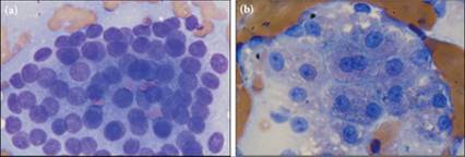
Figures 9.52a,b Large aggregates of intact nuclei in a pool of cytoplasm without distinct borders give a naked-nuclei appearance in this aspirate from a dog. (a) This slide contains essentially only neoplastic cells. (b) A second slide contains clusters of intact hepatocytes and few neoplastic cells to help confirm that the nodule is associated with the liver. Note that several of the hepatocytes in the center of the image are binucleate. Naked nuclei tumors are most commonly of neuroendocrine histogenesis. Distinguishing the tissue of origin of naked nuclei tumors is usually not possible by cytology alone; the combination of history, clinical signs, and special stains is usually needed (Wright–Giemsa, 1,000? magnification).