Mesenchymal tumors
The most commonly reported metastatic mesenchymal tumors include hemangiosarcoma, fibrosarcoma, melanosarcoma, osteosarcoma, and leiomyosarcoma (Figures 9.51a–c; Trigo et al., 1982; Stalker & Hayes, 2007a, b).
Of significant challenge is the fact that, with the exception of melanosarcoma, these tumors can also be primary hepatic mesenchymal tumors. 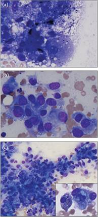
Figures 9.51a–c (a) Most hepatocytes are in poor cytologic condition in this sample. Where visible, there is distinct clear vacuolation consistent with lipid accumulation. (b) Note the spindloid nature of the cells, a cell shape seen with mesenchymal cells, while the pleomorphism suggests a neoplastic process. (c) There are also many pleomorphic mesenchymal cells with erythrophagia, or pale blue cytoplasmic material of unknown significance (a and b, Wright–Giemsa, 1,000? magnification; c, Wright–Giemsa, 500? magnification; inset, Wright–Giemsa, 1,000? magnification).