Epithelial tumors
The presence of nonhepatocellular, distinctly malignant epithelial cells interspersed with normal hepatocytes is evidence of metastatic carcinoma or cholangiocellular carcinoma (Figures 9.6, 9.49a–c, 9.50a–c; Stockhaus et al., 2004).
The most common metastatic carcinomas in the liver include pancreatic, gastric/colonic, renal, mammary, ovarian, and squamous cell carcinomas (Trigo et al., 1982; Stalker & Hayes, 2007a, b). 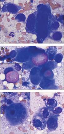
Figures 9.49a–c Liver aspirate from a Beagle. (a) Prominent criteria of malignancy, including anisocytosis, anisokaryosis, a variable N:C ratio, and macro- and micronuclei, are noted. (b) Note the pink cytoplasmic vacuole, which displaces the nucleus, producing a signet ring appearance, in several cells in this image. The inset shows a single intact signet ring cell with a pink cytoplasmic vacuole and ovoid nucleus. Cell-free lipid is also noted as discrete clear spaces in the background outside of the cell. (c) Perinuclear clear vacuolation and micronuclei are present. Similar cells were noted in the sublumbar lymph node of this patient and were interpreted as transitional cell carcinoma or, less likely, squamous cell carcinoma (Wright–Giemsa, 1,000? magnification).
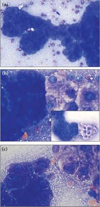
Figures 9.50a–c Canine hepatic aspirate. (a) Many distinct papillary structures are found (Wright–Giemsa, 1,000? magnification). (b) These neoplastic cells (left side of image) strikingly contrast with hepatocytes observed on the right side of the image. The hepatocytes often have cytoplasmic rarefaction consistent with glycogen or hydropic degeneration (main image, Wright–Giemsa, 1,000? magnification; inset, Wright–Giemsa, 500? magnification). (c) Where a thin enough layer of cells is present, the neoplastic cells (left side of image) have deeply basophilic cytoplasm with fine pink granules, prominent cell–cell junctions, cell crowding, multiple nucleoli, and mild anisocytosis and anisokaryosis. In combination, these findings suggest carcinoma. The background is blue–gray flecked with pink granules, similar to the cytoplasm of the neoplastic cells; this feature indicates either necrosis or fragile cells that ruptured during sample preparation. The cells in the upper right are hepatocytes (Wright–Giemsa, 1,000? magnification).