Plasmacytoma/multiple myeloma
Neoplastic populations of plasma cells have been observed in the liver of both dogs and cats, either as a primary mass or as a metastatic lesion. Multiple myeloma and other myeloma-related disorders more in cats often involve the liver and spleen without bone marrow involvement (Mellor et al., 2008).
Cytologically, well-differentiated plasma cells have a moderate amount of basophilic to lightly basophilic cytoplasm, which frequently contains a paranuclear clearing and an eccentrically located nucleus with clumped to smooth chromatin (Figures 9.47a, b). Binucleation and atypia are common features of neoplastic plasma cells. Neoplastic plasma cell morphology can be highly atypical and not look like typical plasma cells, especially in aggressive tumors. (Mellor et al., 2008; Wachowiak et al., 2022).The distinction between solitary plasma cell tumor, multiple myeloma, and other myeloma-related disorders cannot be made solely by cytology; however, cytologic findings in conjunction with radiographic or advanced imaging, serum or urine protein electrophoresis, and immunofixation can provide enough evidence to reach an antemortem diagnosis.
Metastatic neoplasms
The liver is a highly vascularized organ, which receives the first pass at nutrients arriving from the gastrointestinal tract and is the source of much anabolic activity; therefore, it is the ideal metastasis site for many neoplasms. As a general rule, metastatic neoplasms can, but do not necessarily, retain some of the characteristic features of the tissue of origin. Often, close scrutiny will allow diagnosis of a specific metastatic tumor, sometimes diagnosis only to the cell line (epithelial versus mesenchymal) can be achieved, and other times the neoplasm is anaplastic enough that the only accurate cytologic diagnosis available is ‘metastatic neoplasm’ (Figures 9.48a–c). With the increased availability of immunodiagnostics, identification of specific cellular markers should be able to help differentiate more challenging tumors.
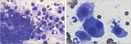
Figures 9.47a,b (a) Large round cells with eccentric nuclei and smooth moderately basophilic cytoplasm are present within this feline hepatic aspirate. Note that many of the neoplastic cells are as large as, or larger than, surrounding hepatocytes and display marked anisocytosis and anisokaryosis (Diff-Quik®, 500? magnification). (b) Occasionally, the neoplastic cells are binucleate. Contrast the size of the neoplastic cells with the size of the neutrophils in the image (Diff-Quik®, 1,000? magnification). This cat had a plasma cell tumor.
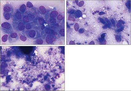
Figures 9.48a–c (a, b) Very few hepatocytes are found in this sample. Instead, abundant anaplastic neoplastic cells, which are found in both cohesive clusters and looser aggregates of spindloid cells, occasionally associated with pink extracellular material, are present. The conflicting cell morphologies prevent cytologic determination of tissue origin. If sufficient tissue architecture is present, the histogenesis of anaplastic tumors may be determined by biopsy and histopathology. (c) Several areas of blue–gray material, consistent with necrosis, are also present (Wright–Giemsa, 1,000? magnification).