Histiocytic sarcoma
Histiocytic sarcoma has been described in the liver of dogs, most commonly as part of disseminated histiocytic sarcoma. However, histiocytic sarcoma rarely presents as a localized tumor found in the limbs or visceral organs, including the liver (Constantino-Casas et al., 2011).
The cells in histiocytic sarcoma have a marked amount of cellular atypia and mitotic activity (Figures 9.46a–d). Most cells have lightly to moderately basophilic and often faintly granular cytoplasm, which contains fine to small punctate vacuoles. The nuclei are round to ovoid to reniform and indented with one to several prominent nucleoli. Marked variation in N:C ratio, anisocytosis, anisokaryosis, bi-, tri-, and multinucleated cells, and many exceptionally large cells are observed. Erythrophagia is another common feature; many patients with histiocytic sarcoma will present with a prominent anemia (Constantino-Casas et al., 2011). Together, these criteria can give the suggestive diagnosis of histiocytic sarcoma; the addition of immunocytochemical staining has been used to confirm the diagnosis (Sapierzyński et al., 2012). Histiocytic sarcoma is a malignant transformation of either dendritic cell lines (Langerhans or interstitial lineages) or macrophages. Therefore, histiocytic sarcoma should be positive for CD1, MHC-II, CD18, and vimentin; negative for CD3, CD79a, PAX5, CD20 and cytokeratin; and may display lineage-dependent markers such as CD11c, CD11d, or E-cadherin (Moore et al., 2006; Constantino-Casas et al., 2011; Moore, 2014). 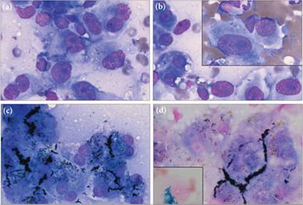
Figures 9.46a–d Histiocytic sarcoma in a Bernese Mountain Dog. (a, b) Many large spindloid to ovoid cells with multiple nucleoli, anisocytosis, and anisokaryosis are found. The overtly neoplastic mesenchymal cells and breed predilection suggest histiocytic sarcoma.
(c) Hepatocytes in this sample had both coarse blue–black intracellular bile casts and cytoplasmic blue–green and pale brown granulation consistent with lipofuscin and bile. (d) Neither the blue–green material nor the brown pigment is positive for iron by the Prussian blue reaction. Inset image is from the control slide; note the blue-staining iron within this macrophage (a–c, Wright–Giemsa, 1,000? magnification; d, Prussian blue, 1,000? magnification).
Disseminated histiocytic sarcoma is found most commonly in Bernese Mountain Dogs, where it has a polygenic mode of inheritance. It is also common in Rottweiler and Retriever breeds and has been diagnosed in multiple other canine breeds (Padgett et al., 1995; Abadie et al., 2009). Flat-coated Retrievers have an increased predilection for the localized form of histiocytic sarcoma (Constantino-Casas et al., 2011).
Histiocytic sarcoma is less commonly observed in cats. Both of the feline-specific histiocytic mass lesions, feline pulmonary Langerhans cell histiocytosis and feline progressive histiocytosis, have been reported to have hepatic involvement. There are very few published reports of these entities, making the description of their epidemiology, clinical progression, and morphologic characteristics uncertain; however, data suggest a poor long-term prognosis (Busch et al., 2008; Moore, 2014).