Mast cell tumor
Mast cells are present in low numbers in most hepatic aspirates and numbers can increase with inflammation and fibrosis; therefore, care should be used when diagnosing a hepatic mast cell tumor (Stockhaus et al., 2004; Masserdotti, 2016).
Mast cells are individualized round cells with distinct metachromatic purple granules, which commonly obscure nuclear morphology when stained with typical Romanowsky stains (Figures 9.16, 9.45a, b). They can occasionally appear agranular if granule content is washed away during processing. High numbers of mast cells, mast cells in clusters, or mast cells with variable granulation within the same sample have been criteria used to diagnose mast cell tumors (Stockhaus et al., 2004). Most hepatic mast cell tumors are associated with disseminated disease; however, primary hepatic mast cell tumors have been diagnosed (Takahashi et al., 2000). 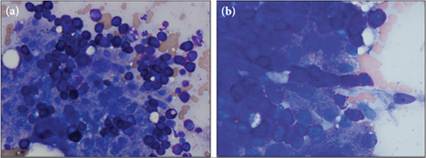
Figures 9.45a,b Liver aspirate from a domestic shorthair cat with mast cell tumors near the anus and within the spleen. (a) Copious mast cells are present throughout the entirety of this sample. Note the pleomorphism and aggregates of mast cells within the hepatic cluster (Wright–Giemsa, 500? magnification). (b) Large aggregates of mast cells with minimal atypia are wedged into the sinusoids between plates of hepatocytes (Wright–Giemsa, 1,000? magnification).
More medical literature on Medic.Studio
More on the topic Mast cell tumor:
-
Infectious diseases -
Internal diseases -
Obstetrics and Gynaecology -
Pediatrics -
Veterinary medicine -
-
Conflictology -
Ecology -
Economy -
Finance -
History -
Law -
Medicine -
Philosophy -
Religious studies -