Nonlymphoid leukemia
Nonlymphoid hematopoietic leukemia has been reported in small animals as both the chronic and acute forms with granulocytic, dendritic, erythroid, and megakaryocytic lineages represented (Figures 9.44a–c; Fine & Tvedten, 1999; Allison et al., 2008; Ameri et al., 2010; Fischer et al., 2012).
As is typical of leukemia, a fairly monomorphic population of hematopoietic cells is observed. If immature forms (blasts) are present, an acute leukemia is diagnosed. Often, bizarre blast cells are present, which prevent determination of the cell line of origin based on Romanowsky stains alone and immunophenotyping is required. With chronic leukemia, a more mature population is present and the cell line of origin is more apparent. 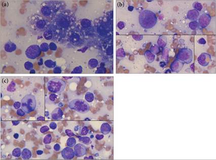
Figures 9.44a–c Liver FNA from an 11-year-old Rottweiler. (a) Very few clusters of hepatocytes are present in this aspirate; however, there are many individualized round cells. (b) These cells have a minimal amount of deeply basophilic cytoplasm and single round nucleus, which occasionally is indented. Most cells have few to many pink to azurophilic granules. Note the erythrophagic cell in the lower right corner. (c) Mitotic figures are easily found. A bizarre mitotic figure is in the upper left corner. The large cell size, granulation, and nuclear morphology are consistent with a hematopoietic neoplasm and suggestive of a myeloid lineage; however, histopathology on the liver failed to confirm a neoplastic presence (Wright–Giemsa, 1,000? magnification).
More on the topic Nonlymphoid leukemia:
- Nonlymphoid leukemia
- References
- Hematopoietic abnormalities
- Barger A.M., MacNeill A.L. (Eds.). Small Animal Cytologic Diagnosis: Canine and Feline Disease. CRC Press,2024. — 536 p., 2024
- Chronic Allograft Dysfunction
- Slide preparation
- Malignant transformation: a multistep process
- Microbiology
- Sarcoma
- Macrovascular Complications of Diabetes Mellitus