Slide preparation
Slides should be prepared immediately after sample collection. A drop of marrow is expelled onto one end of a slide, the slide is tilted to let blood run off onto absorbent paper (marrow particles will adhere to the glass slide), and then a second slide is backed into the remaining marrow drop and pulled forward at approximately a 45° angle, analogous to preparing a feather-edge blood film.
In addition to preparing feather-edge type blood films where marrow particles are maintained aggregated, several ‘squash’ type of films should also be prepared by gently dropping a second slide perpendicular to the first slide on the drop of marrow remaining after blood has run off, and pulling that slide forward. In this type of smear, particles are spread apart to facilitate individual cell recognition (Figure 19.7). Two to three of each type of smear should be prepared and rapidly air-dried, either with the aid of a small fan or by vigorously waving slides in the air. Removing blood prior to preparing smears concentrates hematopoietic elements and hastens slide drying, thereby improving cell preservation. It is difficult to impossible to prepare good films from clotted samples though clots may be fixed in formalin for subsequent paraffin embedding and sectioning. 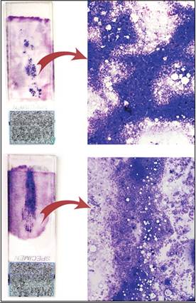
Figure 19.7 Marrow aspirate films with poorly spread dense particles (top) and well-spread particles (bottom). Images on right at 50? magnification.
Alternatively to immediate film preparation, aspirated marrow can be placed in a glass or Petri dish that is then tilted to separate the pale yellowish marrow particles from blood. Particles are then retrieved using a pipette, microhematocrit tube, or hypodermic needle, placed on a slide, and films are prepared as above. Aspirated marrow remaining in the syringe should be placed in a 3 mL EDTA tube and submitted along with the slides in case additional slides need to be prepared or special tests are anticipated.
Normal marrow aspirates have cell concentrations >50,000/μL; therefore, even a small volume of marrow can yield abundant cells for flow cytometry, immunochemistry, or assessment by PCR (Tan et al., 2014a).If there is doubt about the quality of the marrow aspirate, or particles are not grossly visible, it is advisable to stain one slide with a quick-type Romanowsky stain (e.g. Dip Quick ®, Jorvet) before recovering the patient from general anesthesia. A good quality marrow film should have grossly apparent granular material and cell-dense areas admixed with blood (Figure 19.8). Microscopically, there should be abundant particles and hematopoietic cells. If slides are not cellular, aspiration should be repeated. Unsuccessful aspiration or poorly cellular samples are not necessarily a result of poor technique but are more likely to reflect marrow diseases such as myelofibrosis, myelophthisis, or aplastic anemia (Donald & Kakkar, 2021). In these cases, marrow core biopsy is required for diagnosis.
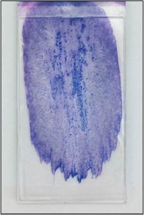
Figure 19.8 Example of well-prepared BM aspirate smear, showing darker purple BM particles surrounded by blood.
Whole anti-coagulated marrow can be assessed in automated hematology analyzers if grossly visible clots are first manually removed (for example with wooden applicator sticks). Analysis of nucleated cells by size and peroxidase content in instruments such as the Advia 2120 (Siemens) or by light scatter and fluorescence in the Sysmex XT2000iV yields rapid information about sample cellularity, cell types, reticulocyte concentration, and cell distribution (Figure 19.9, Tan et al., 2014a; Criswell et al., 2018; Pernecker et al., 2017).
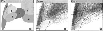
Figures 19.9a–c Analysis of marrow aspirates in the automated hematology analyzer Advia 2120 yields differential cell estimates. (a) 0 = Cells lacking peroxidase activity are lymphocytes and large unstained cells (LUC); 1 and 2 = large cells with low peroxidase activity are immature granulocytic cells; 3 = medium-size cells with low to moderate peroxidase activity are monocytic cells; 4 = medium-size cells with moderate to high peroxidase activity are likely metamyelocytes; 5 = large cells with high peroxidase activity are likely band neutrophils; 6 = medium-size cells with high peroxidase activity are likely segmented neutrophils. (b) Granulocytic hyperplasia with numerous mature granulocytes. (c) Acute myeloid leukemia with partial neutrophil differentiation.
More on the topic Slide preparation:
- Slide preparation
- Staining of slides
- Cytologic evaluation
- Sampling
- Slides for cytologic evaluation
- General Principles of Histology
- Barger A.M., MacNeill A.L. (Eds.). Small Animal Cytologic Diagnosis: Canine and Feline Disease. CRC Press,2024. — 536 p., 2024
- The Use of the Microscope
- Urinary sediment
- Impression smears