Cytologic evaluation
Once the slide is stained, it is ready to be evaluated cytologically. Low-power microscopic evaluation is critical. Many different patterns, processes, and background material can be very easily identified with low-power evaluation.
Generally, for cytology, this is done at 10? magnification; however, visual evaluation of the stained slide itself is also beneficial to determine the position of the sample on the slide (Figure 1.42). Microscopic low-power evaluation will allow the evaluator to determine if the sample is cellular enough for diagnostic quality. Patterns of cellular association can be identified. Epithelial cells will commonly form clusters whereas round cells and mesenchymal cells are more likely to be arranged in a loose sheet or as individualized cells. Low-power evaluation can also allow for evaluation of the diversity of a population. Recognition of one cell population versus a pleocellular population can help determine the overall process. One cell type being present is more consistent with aspiration of a tumor or of a specific tissue (for example, aspiration of the liver may result in the identification of primarily hepatocytes), while a mixed or pleomorphic population of cells is more consistent with inflammation (Figure 1.43). Low-power evaluation should also be used to identify well-stained, diagnostic areas on the slide. Thick areas of the smear make full evaluation of cellular morphology difficult. Thinner areas, where cells have the opportunity to spread out, are more useful for evaluating individual cellular morphology (Figures 1.44a, b). 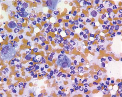
Figure 1.43 FNA from a dog reveals a mixed cellular population consistent with an inflammatory response (Wright–Giemsa, 500? magnification).
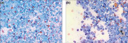
Figures 1.44 Lymph node aspirate from a dog.
(a) Reveals a thick area of the smear. The cells are crowded and inappropriately stained. (b) Represents a thinner area of the smear. The cells are more spread out and appropriately stained (Wright–Giemsa, 500? magnification).
At higher magnification, individual cellular features, such as chromatin pattern, presence of nucleoli, and cytoplasmic features, can be identified as well as smaller microorganisms such as bacteria (Figure 1.45). It is important to clearly identify the presence of cytoplasm and a nucleus in the cells. Cells may rupture during the aspiration or slide preparation process, resulting in bare nuclei or streams of nucleoproteinaceous debris (Figure 1.46). Nuclei that have lost their cytoplasm will swell and become much larger, and it is important not to overinterpret their value. A diagnosis should always be made on intact cells.
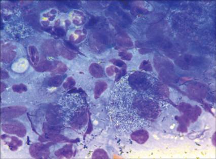
Figure 1.45 Lymph node aspirate from a dog. At higher magnification, macrophages containing negative staining rod-shaped bacteria (Mycobacteria sp.) can be more easily identified (Wright–Giemsa, 1,000? magnification).
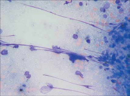
Figure 1.46 FNA from a mass on a dog. Streams of nucleoproteinaceous debris can be identified and are often caused during slide preparation. These structures should not be confused with fungal hyphae (Wright–Giemsa, 500? magnification).
Higher magnifications can include 40?, 50?, and 100? objective lenses. Often 100? is necessary to fully evaluate the sample for bacteria and smaller yeast. If using a 40? objective that is not oil immersion, remember to place a coverslip over the sample to improve the crispness of the objective. Features of individual cells will be described in each of the chapters.
Finally, the material present in the background should be examined.
Blood contamination is possible with every aspirate, although some organs such as the spleen and liver are more vascular than others and are prone to lead to significant hemodilution when sampled. Accompanying white blood cells can be seen in samples with significant peripheral blood contamination; therefore care should be taken not to confuse inflammation with blood contamination. Proteinaceous material can also be observed, either as a pathologic process or potentially as a healthy cellular product, such as mucin production in joint aspirates. If the sample is highly proteinaceous, the individual cellular features may be difficult to evaluate, so identification of a thin area in the smear is recommended (Figure 1.47). Thick eosinophilic-staining proteinaceous material may indicate the presence of osteoid, chondroid, fibrin, or collagen in a sample. Collagen may be seen accompanying mast cell tumors or epithelial neoplasms (Figure 1.48). Granules can also be seen in the background from ruptured melanocytes, mast cells, eosinophils, and, less likely, basophils. It is important not to confuse these structures with microorganisms. Again, it is important to try to identify the intact cells and compare the background material with any granules in the cytoplasm of the cell population (Figure 1.49). 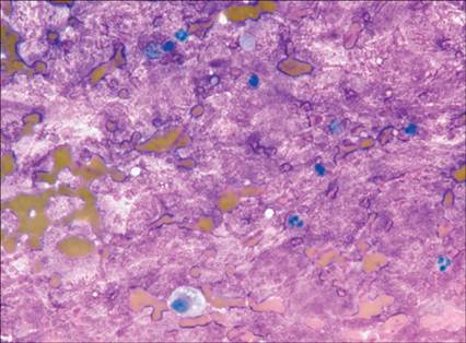
Figure 1.47 Aspirate from a lytic bone lesion in a dog. The deeply eosinophilic material in the background is consistent with chondroid. The material is so thick, it is difficult to fully evaluate the nucleated cells in the sample (Wright–Giemsa, 500? magnification).
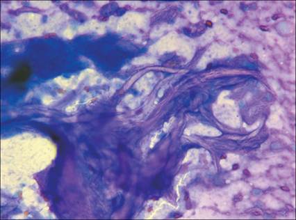
Figure 1.48 Aspirate from a mast cell tumor in a dog. Thick aggregates of collagen bundles are present (Wright–Giemsa, 500? magnification).
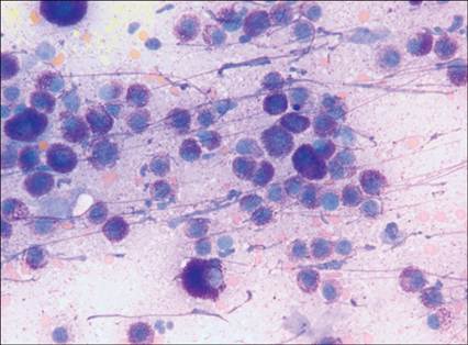
Figure 1.49 Aspirate from a mast cell tumor in a dog.
Metachromatic granules are visible in the cytoplasm of the cells but also in the background (Wright–Giemsa, 500? magnification).
In order to interpret the cytology appropriately, it is important to consider the histologic appearance of the tissue and the underlying architecture. The term exfoliate cytology refers to looking at individual cells been exfoliated from a tissue – literally the tissue has been ‘stripped of leaves’. In order to accurately interpret the cytologic findings, it is essential to know the structure of the ‘tree’ from which the cells originate. There is a close relationship between cytologic findings and the histologic structure of common tissues and tumors. One frustration for the beginner cytologist is the question of identifying normal, particularly as normal structures are rarely intentionally aspirated. For those without a strong background in histology, a good reference text in the clinic can aid in the recollection of which types of cells are normally found in each tissue. This is particularly helpful in the identification of cell types in aspirates from unusual locations, such as the esophagus or reproductive organs. Also bear in mind that there are some species differences in what is normal in various locations (Bacha & Bacha, 2012).
A recent article highlighted common patterns in cytologic samples and stressed the importance of careful attention to collection and smear preparation (Masserodotti, 2006). Patterns are found more easily if the relationships among cells are maintained and cells are intact. These patterns correlate to the parent tissue architecture and can be explained by the normal function of the tissue.
Pavement pattern
Pavement patterns are found in squamous, conjunctival, and urogenital epithelium. These cells often exfoliate in loose cohesive sheets or individualized cells. They tend to be flat with somewhat angular margins. This pattern is often confused with individual spindle cells because of the angular cell margins and poor cohesion (Figure 1.50).
Additionally, many of these tissues have multiple layers of cells with distinct morphology that may be aspirated in the same cytologic sample. The prime example of this is the presence of variable maturing cells in cystic skin tumors. The cells range from mature anuclear keratin flakes to squamous cells with nuclei in various stages of pyknosis (Figure 1.51). The architecture of the various benign skin tumors explains the variability of the cystic epidermal neoplasms seen on cytology (Gross, 2006). 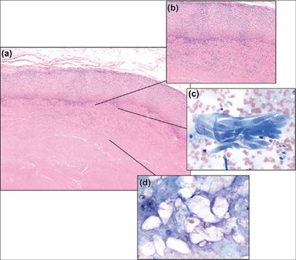
Figure 1.51 Histologic (a, 100? magnification; b, 400? magnification) and cytologic (c & d, 500? magnification) examples of a type of cystic skin tumor (pilomatricoma) from a cat. The epithelial cells (c) form a flattened pavement pattern on the perimeter while the center of the cyst (d) contains cholesterol crystals and poorly staining keratin debris (H&E and Wright–Giemsa).
Honeycomb and palisade patterns
The honeycomb and palisade patterns are related to the pavement pattern; however, they tend to maintain a more cohesive appearance. These are cuboidal or columnar cells that maintain distinct cell margins and cohesiveness. The most common site in which to observe the honeycomb pattern in veterinary cytologic samples is the normal prostate or benign prostatic hyperplasia (Figure 1.52). It can also be seen in the normal stomach and intestine; however, these tissues are less commonly sampled for cytology. The palisade pattern presents as ribbons and small aggregates of cuboidal to elongated cells and is common in benign skin tumors (Figures 1.53, 1.54).
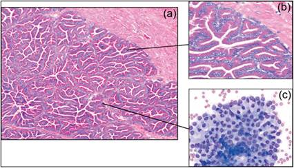
Figure 1.52 Histologic (a, 100? magnification; b, 400? magnification) and cytologic (c, 500? magnification) examples of benign prostatic hyperplasia in a dog. The cells exhibit a classic honeycomb pattern.
They are columnar cells with basal nuclei and a moderate amount of cytoplasm, which stains eosinophilic on histology and basophilic on the cytology preparation (H&E and Wright–Giemsa).
alt=fig1.53.jpg>
Figure 1.53 Histologic (a, 200? magnification; b, 400? magnification) and cytologic (c, 500? magnification) examples of trichoblastoma from a dog. This is one of several different skin tumors that are classified as basaloid epithelial tumors on cytology as they exfoliate palisading ribbons of cuboidal cells. Collagen and fibrocytes in surrounding matrix (arrowheads) can be seen on the histology slides but not on the cytology preparation (H&E and Wright–Giemsa).
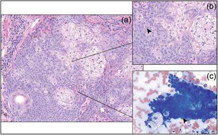
Figure 1.54 Histologic (a, 100? magnification; b, 400? magnification) and cytologic (c, 500? magnification) examples of a sebaceous epithelioma from a dog. Dense cuboidal cells and large foamy sebaceous cells can be seen on both preparations. Arrowheads (b, c) indicate the cells in a palisade pattern similar to those seen in Figure 1.53 (H&E and Wright–Giemsa).
Acinar pattern
The acinar pattern reflects the secretory nature of glandular epithelium. When the cells exfoliate intact, they are arranged around an empty or secretion-filled central area. Many of these tissues are prone to mechanical disruption, resulting in loosely cohesive sheets or nuclei floating in a ‘sea of cytoplasm’. Thyroid carcinomas and anal gland carcinomas are known for this type of presentation (Figure 1.55).
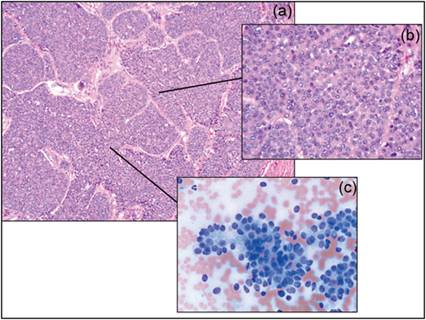
Figure 1.55 Histologic (a, 100? magnification; b, 400? magnification) and cytologic (c, 500? magnification) examples of an apocrine anal gland adenocarcinoma from a dog. The acinar arrangement can be seen on the edge of the sheets of loosely cohesive epithelial cells (H&E and Wright–Giemsa).
Supporting stroma
Mesenchymal cells create the supportive framework of all tissues. They provide a tight interwoven matrix, which is more difficult to aspirate and thus typically yields fewer cellular samples. Normal fibrocytes can be seen as the pink bundles between the cellular islands of the trichoblastoma in the histopathology sample of Figure 1.53 but they were not present in the cytology sample from that tumor. Abnormal mesenchymal populations such as tumors or granulation tissue will exfoliate more readily than normal fibrous tissue, and the resulting slides have increased cellularity. They present as individual spindle cells in a storiform pattern (Figure 1.56). In some cases, the vascular elements of a tissue will be present on a cytology sample. These small capillaries are commonly seen in thicker preparations of some soft tissue sarcomas (Figure 1.57). In addition, the spindle cells may be associated with extracellular stromal elements such as chondroid or osteoid, which have an amorphous purple–pink appearance (Figure 1.58).
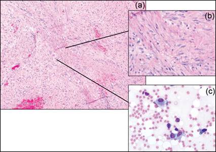
Figure 1.56 Histologic (a, 100? magnification; b, 400? magnification) and cytologic (c, 500? magnification) examples of granulation tissue from a healing surgical site in a dog. The individual spindle cells seen in the cytology panel are difficult to distinguish from a well-differentiated sarcoma (H&E and Wright–Giemsa).
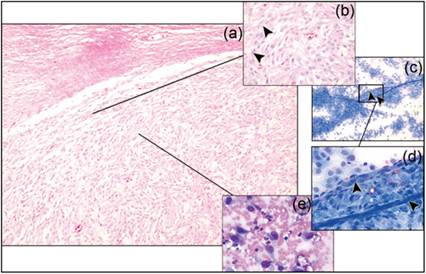
Figure 1.57 Histologic (a, 100? magnification; b, 400? magnification) and cytologic (c, 100? magnification; d & e, 500? magnification) examples of a soft tissue sarcoma from a dog. The spindle cells are seen both individually and in a storiform arrangement near capillaries. Arrowheads (b, c, d): capillaries (H&E and Wright–Giemsa).
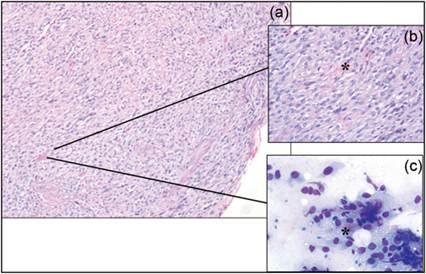
Figure 1.58 Histologic (a, 100? magnification; b, 400? magnification) and cytologic (c, 500? magnification) examples of an osteosarcoma from a dog. The asterisks (b, c) represent osteoid seen as a pink matrix material on both stains (H&E and Wright–Giemsa).
Complex tissues
Samples such as internal organs, inflammatory samples, and tumors with multiple tissue types can lead to confusing cytologic samples. The normal architecture and proportion of different cell types should correspond to what exfoliates on cytology. Mammary tumors are a prime example of tissues that yield multiple cell types on cytology. They often have a mixture of glandular epithelial cells and mesenchymal cells. These mesenchymal cells can range from spindeloid fibrocytes to chondrocytes and osteoblasts if the tumor contains complex features such as cartilage or bone (Figure 1.59). Some tumors, such as the perianal gland adenoma in dogs, have both large polygonal epithelial cells and smaller cuboidal reserve cells. Understanding the histology helps explain the presence of these two cell types (Figure 1.60).
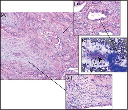
Figure 1.59 Histologic (a, 100? magnification; b & d, 400? magnification) and cytologic (c, 500? magnification) examples of a complex mammary tumor from a dog showing both epithelial and mesenchymal differentiation. The spindle cells are intermixed with extracellular matrix material consistent with osteoid (arrowhead, c) and are adjacent to cuboidal cells in a palisade pattern (H&E and Wright–Giemsa).
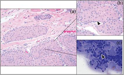
Figure 1.60 Histologic (a, 100? magnification; b, 400? magnification) and cytologic (c, 500? magnification) examples of a perianal adenoma from a dog. Arrowheads (b, c): cuboidal reserve cells (H&E and Wright–Giemsa).
The liver is another tissue in which multiple cell types are normal. The majority of the cells seen on cytology should be hepatocellular, with a smaller proportion of biliary epithelial cells. Inflammatory cells can be diffuse but are often overrepresented in the periportal area. An example of a liver from a cat with histoplasmosis is shown (Figure 1.61).
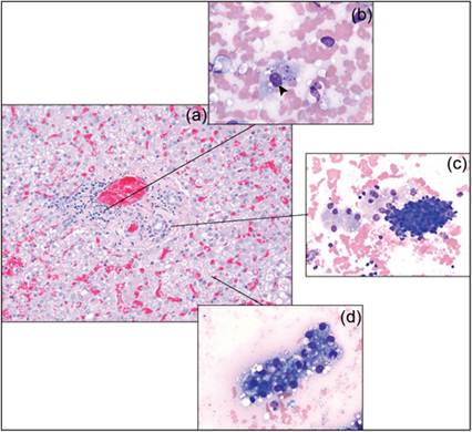
Figure 1.61 Histologic (a, 400? magnification) and cytologic (b, c & d, 500? magnification) examples of a liver. Arrowhead (b): macrophage containing Histoplasma capsulatum yeast. Macrophages and biliary epithelial cells (c) are mixed with vacuolated hepatocytes (d) (H&E and Wright–Giemsa).
Inflammatory lesions can have a myriad of cell types present. The cells seen may not reflect the underlying pathology in the case of a tumor with a devitalized center or when the inflammation is incidental to the final diagnosis. As seen in Figure 1.56, reactive spindle cells can mimic neoplastic processes and are often intermixed with inflammatory lesions. An example of the drastic difference inflammation can make in your ability to diagnose simple lesions on cytology is when there is granulomatous inflammation in a simple lipoma. Fat has a characteristic cytologic appearance (Figure 1.62), but when traumatized it produces an intense granulomatous steatitis. Aspiration of this inflammatory population can be misinterpreted as a primary inflammatory or even a neoplastic process such as a histiocytic sarcoma (Figure 1.63).
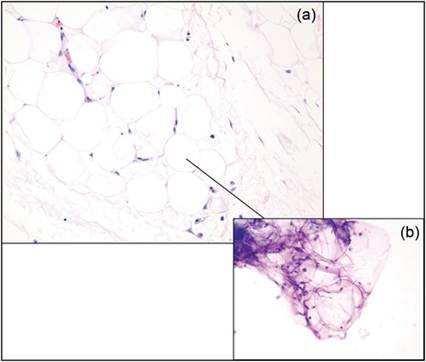
Figure 1.62 Histologic (a, 200? magnification) and cytologic (b, 200? magnification) examples of a lipoma. The fat-filled cells are similar in both preparations (H&E and Wright–Giemsa).
alt=fig1.63.jpg>
Figure 1.63 Histologic (a, 200? magnification; b, 400? magnification) and cytologic (c, 500? magnification) examples of an infiltrating lipoma with inflammation. The majority of cells in this cytology preparation are large activated macrophages and non-degenerate neutrophils. There is a background of fatty material but only rare intact adipocytes (H&E and Wright–Giemsa).
A normal salivary gland has a characteristic appearance of foamy epithelial cells in small sheets, but when there is inflammation and dilation of the salivary duct, a sialocele may form, producing a marked granulomatous inflammation (Figure 1.64).
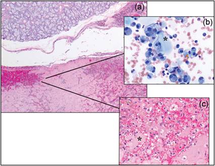
Figure 1.64 Histologic (a, 200? magnification; c, 400? magnification) and cytologic (b, 500? magnification) examples of a sialocele. The normal gland can be seen above the dilated sialocele. The aspirate (b) revealed abundant vacuolated macrophages, some of which contained hematoidin crystals. The blue inspissated saliva on the cytology (asterisk, b, c) stains pink in the histology section (H&E and Wright–Giemsa).
A case that caused a diagnostic dilemma serves as the final example to instill caution in the overzealous cytologist (Figure 1.65). The spindle cells and multinucleate cells were the majority of the cells on the slide; there was relatively little keratin debris and inflammation, and a sarcoma was suspected. Histopathology revealed the lesion to be a benign cystic skin tumor with inflammation. This example highlights the need for caution in the interpretation of cytologic samples without the correlation of the tissue architecture.
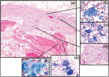
Figure 1.65 Histologic (a, 100? magnification; d, 400? magnification) and cytologic (b, c, e & f, 500? magnification) samples from a cat skin tumor. The majority of the cells on this cytology were the spindle cells and multinucleate cells seen in c and e. The inflammatory cells in b and the keratin debris in f were rare on the cytology slide. Biopsy revealed a keratin-filled cyst with a border of multinucleate inflammatory cells, fibrosis, and inflammation (H&E and Wright–Giemsa).