Staining of slides
If sending slides to a laboratory, it is recommended that unstained slides be submitted. It is beneficial, however, to examine at least one slide to determine if an adequate sample was obtained and/or the appropriate tissue sampled (Figure 1.38).
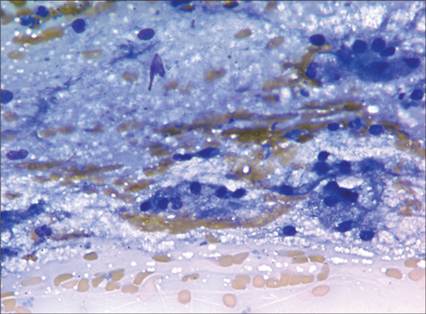
Figure 1.38 Mandibular lymph node aspirate from a dog reveals cytologically unremarkable salivary tissue. Clusters of vacuolated salivary epithelial cells and thick mucus are observed in the background. No lymphoid tissue is observed, so this is likely inadvertent aspiration of healthy salivary tissue (Wright–Giemsa, 1,000? magnification).
Many types of stains are available for cytologic preparations, and some are available for easy in-house use. Common types used in veterinary medicine include Romanowsky stains, Pap stain, and new methylene blue. Each of these will be described in more detail.
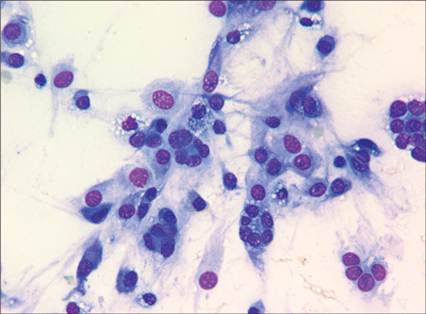
Figure 1.39 Subcutaneous mass on the carpus of a dog. The sample is highly cellular and consists of a population of neoplastic mesenchymal cells. The sample is adequately stained with basophilic cytoplasm, classic purple staining of the nucleus and basophilic nucleoli (Wright–Giemsa, 1,000? magnification).
Romanowsky stains
In veterinary medicine, Romanowsky stains are the most commonly used and readily available stains. These stains include May–Grünwald–Giemsa, Leishman–Giemsa, Wright–Giemsa, and Diff-Quik® and its variants. The Romanowsky stains include a combination of reagents that include azure B/polychromed methylene blue and the eosin family of stains (Horobin, 2011). The characteristic staining with this combination results in a classic purple staining of the nucleus (Figure 1.39).
If the nucleus of the cell is not purple, concern for inappropriate staining, inadequately dried sample, overly thick preparation, or exposure to aldehyde fixatives should be considered (Horobin, 2011; Kraft's, 2011).One of the many advantages of Romanowsky stains is the ease of fixation. Air drying is all that is required. Even heat fixation is unnecessary for these types of stains. A recent study showed that even for ear swabs, heat fixation provided no benefit (Toma et al., 2006). Slide preparation, therefore, is so important because quick, even drying is essential for uniform staining. Prolongation of the drying time can result in cellular rupture and poor cell preservation (Jorundsson et al., 1999). Additionally, fixation will influence the uptake of the stain. The result of an inappropriately fixed slide is poorly stained cells with blue rather than purple nuclei (Horobin, 2011). Incomplete staining or understaining of samples is a common problem. As mentioned earlier, the nuclei of cells stained with Romanowsky stains should be purple. If the nuclei are blue, the sample may have not stained long enough, had prolonged drying time, be too thick, or been exposed to aldehyde fixatives (Figures 1.40a, b). A blue–green appearance to the erythrocytes indicates the sample has been exposed to aldehyde fixatives or the pH of the stain is too high (Horobin, 2011).
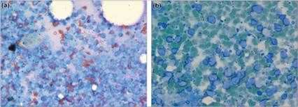
Figure 1.40a,b Lymph node aspirate from a dog with lymphoma. (a) Represents a sample that is poorly stained. The nuclei are staining blue rather than purple and individual cytologic features are difficult to discern. (b) Represents a sample exposed to formalin fumes. Note the green staining of the erythrocytes and the blue staining of the nuclei (Wright–Giemsa: a, 500? magnification; b, 1,000? magnification).
There are several different Romanowsky stains available. Many practices will use a manual stain like Diff-Quik®.
The manual stains are presented as three solutions: methanol, solution 1 or eosin, and solution 2 consisting of azure A and B. The slides are dipped in each solution. This is an easy stain to use; however, there are some limitations. The solutions must be changed frequently to limit sedimentation and bacterial growth. Additionally, mast cell granules, basophils, and granules from granular lymphocytes do not stain as reliably with the aqueous Romanowsky stains (Allison & Velguth, 2010). For this reason, many clinical pathology laboratories use Wright–Giemsa or May–Grünwald–Giemsa because the Giemsa component stains mast cell granules vibrantly. If performing in-house cytology, it is important to leave some of the slides unstained so one of these stains can be used.There are several advantages to the use of Romanowsky stains for cytology, including detailed staining of the cytoplasm, background material, and microorganisms (Kraft's, 2011). A more complete list is shown in Table 1.1.
Papanicolaou stain
The Papanicolaou (Pap) stain has a long history of use in cytology. It was developed by George Papanikolaou and is most commonly associated with the Pap smear used to diagnose cervical cancer. The classic stain involves a combination of five dyes including hematoxylin, orange G, and eosin 50, which is a combination of eosin, Bismark brown, and light green. The eosin gives a pink color to the cytoplasm of mature squamous epithelial cells, nucleoli, and cilia. Light green stains the cytoplasm of metabolically active cells blue, such as parabasal cells, squamous epithelial cells, and intermediate and columnar epithelial cells. Hematoxylin is used to stain nuclei (Perez et al., 2005).
The original Pap stain is a little cumbersome for in-house cytology, requiring many reagents and steps. However, simplified Pap stains have been evaluated for easy cytologic use. Sawa et al. (2012) evaluated RADPap® and found the stain was easy to perform and only took 15 minutes; however, there were still 18 steps, which diminishes the clinical utility.
The Ultrafast Papanicololaou staining protocol has 14 steps but only takes 5 minutes to perform (Perez et al., 2005).The Pap stain requires wet fixation such as 95% ethanol, 100% ethanol, or 80% isopropanol (Jorundson et al., 1999). To improve the clinical utility of wet fixation, techniques have also been developed for air-dried smears to be rehydrated with saline (Jorundson et al., 1999).
There are significant advantages of the Pap stain when compared with Romanowsky stains in exhibiting nuclear detail and for evaluating thick tissue samples (Table 1.1; Jorundson et al., 1999; Krafts, 2011).
Table 1.1 Advantages and limitations of the common stains used for cytologic preparations
| Romanowsky | Papanicolaou | New methylene blue | |
| Advantages | Excellent cytoplasmic detail. Adequate staining of background material. | Excellent nuclear detail. Useful in staining cells in thick preparations. | Consistent staining of mast cell granules. Nuclear detail is adequate. Easy to use. |
| Limitations | Staining of cells challenging in thick samples. Nuclear detail is minimal. | Wet fixation or rehydration of samples required. Not useful for evaluation of microorganisms or background material. | Use of stain on air-dried smears for cytology is not permanent. |
New methylene blue
New methylene blue (NMB) is a supravital stain and is very easy to use. It is commonly used to quantify reticulocytes in the peripheral blood because it will stain organelles vibrantly. Its use in cytologic preparations is a little different. A drop of stain is placed on an air-dried smear and then a coverslip is placed on the droplet to evenly spread the stain on the smear (Figures 1.41a–c). This stain provides excellent nuclear and nucleolar detail and also stains mast cell granules (Table 1.1). The stain is a wet mount stain so it is not permanent.
As the NMB stain dries, the cellular staining diminishes. Therefore, this may be a limitation for its use in laboratories required to save slides for an extended period of time. 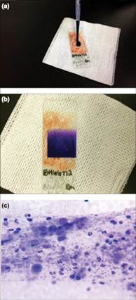
Figure 1.41 New methylene blue staining of cytology specimens. (a) The stain is applied to an unstained cytologic specimen. (b) A coverslip is placed on the slide. (c) Microscopic evaluation reveals reasonable nuclear staining; neutrophils and macrophages are identified in this sample (500? magnification).
Additional techniques for immunostaining
Immunocytochemical staining can be performed on regular unstained or destained cytology preparations. Formalin fixation and antigen retrieval are not required. A limitation of this is the variability in cellularity and cellular preservation. There are some interesting techniques available to improve the consistency of cellularity, but these require formalin fixation and antigen retrieval. Needle rinse cell blocks can be used to prepare cells obtained with cytology for paraffin embedding. These samples are fixed and undergo similar processing as histopathology samples (Marrinhas et al., 2021). The advantage of this technique is to get multiple uniform slides for evaluation, and multiple stains can be performed if needed. An additional technique of placing the aspirate directly in formalin and using a micromesh strainer to place the material in a cassette for processing and paraffin embedding has also been evaluated (Etzioni, 2022). This cyto-histo technique can be useful for immunostaining.
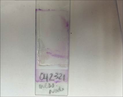
Figure 1.42 FNA from a mass on a dog. Macroscopic evaluation of the slide is beneficial to identify the best area to evaluate. Some aspirates provide a very small sample.