Slide submission
Labeling of the slides is an invaluable component of slide submission whether the slides are being sent out to a clinical pathology laboratory or being evaluated in-house. Pertinent information to write on the slides includes the patient’s identification information (either name or medical record number) and the site sampled, particularly if multiple sites were sampled (Figure 1.34).
Slides with a frosted edge are ideal because then the slides can be labeled with pencil. Special markers or diamond-etched pencils are necessary to label slides without frosted edges (Figure 1.35). Ink from other markers, such as Sharpie® markers, will wash off during the staining process. 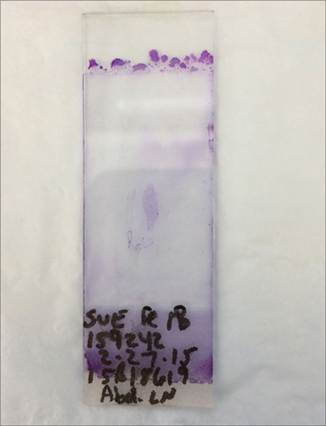
Figure 1.34 FNA from a cat. The slide is labeled with the patient’s name, medical record number, date, and source of the aspirate.
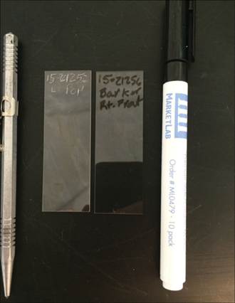
Figure 1.35 Diamond tipped pencil (left) and slide marker (right) used for permanent labeling of samples.
If sending slides to a laboratory for evaluation, it is important to package the slides appropriately because they are quite fragile. Many types of slide boxes are available and include cardboard, polystyrene, and plastic (Figure 1.36). Cardboard carriers are the least expensive; however, they are also the least stable (Figure 1.37). The plastic containers are the most stable, but even these should be wrapped in additional padding or sent in a padded envelope. Accompanying the slides should be a form containing necessary patient information including signalment and pertinent historical data as well as the source(s) of the aspirates. This information is vital for the clinical pathologist to be able to provide the most complete cytologic diagnosis, as well as a thorough list of differential diagnoses and suggestions for appropriate additional diagnostic testing.
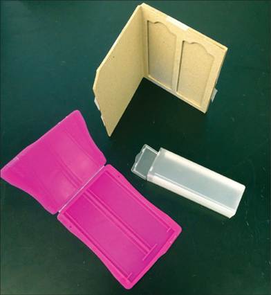
Figure 1.36 Options for containers for slide transport. Plastic slide boxes are more durable than cardboard.
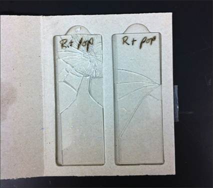
Figure 1.37 Slides sent in a cardboard container, inappropriately packaged. Both slides are severely damaged.