Sampling of intra-abdominal or intrathoracic fluids
Fluid analysis with cytology can be extremely helpful in identifying processes leading to fluid development, including infectious and neoplastic causes. Thoracocentesis and abdominocentesis are fast procedures with a very low risk of complications.
For either body cavity, if a moderate to large amount of fluid is present, it is easy to obtain a sample blindly. Equipment needed includes a 3/4 inch 22-gauge needle, a 3–6 ml syringe, and red and purple top tubes. Unlike when sampling tissues, the needle is always placed on the syringe for this procedure.For abdominocentesis, similar to the standard procedure for cystocentesis, the skin is not usually clipped or scrubbed. To avoid the liver and spleen, the sampling site should be mid-abdomen and towards the right side of the midline. In an upright patient, the needle is inserted into the ventral abdomen, as fluid will sink (Figure 1.32). If the patient is recumbent, the needle is directed into the cavity from below the midline point. The plunger on the syringe is then withdrawn and the sample obtained (Figure 1.33). Several milliliters of fluid can be obtained rapidly. Pressure on the syringe is released before the needle is extracted. The sample is then divided between red and purple top tubes.
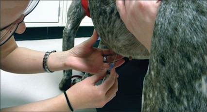
Figure 1.32 Abdominocentesis using a 22-gauge 1½ inch needle and a 6 ml syringe.
alt=fig1.33.jpg>
Figure 1.33 The fluid easily obtained from the abdomen of a dog with marked effusion.
For thoracocentesis, the best place to sample is between the 6th and 8th ribs. The patient is placed in ventral recumbency or may be left standing but restrained. The skin is usually clipped and scrubbed. The needle is inserted between the ribs and ventrally on the chest wall. Depending on the size of the animal, only half the length of the needle may be needed to reach the fluid. Alternatively, an intravascular or butterfly catheter can be used. As with abdominocentesis, the plunger is drawn back and fluid is obtained; pressure on the plunger is released and the needle is withdrawn. For situations where minimal fluid is present in either body cavity, ultrasound guidance can be used to help direct the needle into a pocket of fluid.