Impression smears
Cytologic assessment of tissues that have been biopsied can be a worthwhile diagnostic tool for several reasons. The turnaround time for results is much quicker for cytology versus histopathology, and such results may be critical to guide decisions in time-sensitive situations.
Additionally, some diagnoses can be made more readily with cytology compared with histopathology, including the diagnosis of round cell tumors and mycobacterial infections. Preparing slides for possible cytologic evaluation is a quick and easy procedure to do after a tissue sample has been obtained. Even if the slides are not submitted immediately to save cost when a biopsy is going to be submitted, it is worthwhile saving the slides for later submission if the histopathology suggests that cytology could be of additional advantage.To make an impression smear, the tissue of interest is blotted with gauze to remove surface blood (Figure 1.28). The cut surface of the tissue can then be pressed, quite firmly, to a slide (Figure 1.29). Multiple impressions are made along the length of the slide, and multiple slides are created in this way. For very firm masses, a scalpel blade can be used to scrape at the cut surface in an effort to increase exfoliation (Figure 1.30). Any material on the blade can be smeared onto a slide, and the surface of the tissue itself can again be pressed against slides. When stained, the impression smears will look similar to squash prep samples, with thick and thin areas (Figure 1.31).
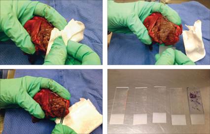
Figures 1.28–1.31 Preparing a surgically excised mass to obtain an impression smear. (1.28) First, gauze is used to dry blood from the surface. (1.29) A slide is pressed firmly against the surface of the mass. (1.30) A scalpel blade is used to scrape cells from the mass; the material on the blade will then be smeared on a slide. (1.31) Impression smear slides, both unstained and stained, showing thick and thin regions.
It is important to remember that the presence of formalin near cytology slides will fix the slides and ruin them for evaluation. Be sure to keep the slides away from the formalin jar, even when it is closed. Histopathology and cytology samples should not be submitted in the same package. Also, if the biopsy samples are very small, care must be taken not to destroy the sample for histologic evaluation. Care and gentle pressure are needed when making impressions of small pieces of tissue, and if multiple biopsies of the same lesion are obtained, only use one or two for impressions.
More on the topic Impression smears:
- Barger A.M., MacNeill A.L. (Eds.). Small Animal Cytologic Diagnosis: Canine and Feline Disease. CRC Press,2024. — 536 p., 2024
- Clostridium piliforme Infection: Tyzzer’s Disease
- Chapter 15 Common gynaecological procedures and surgery
- Chapter 1 The Woman’s Health Examination and Women’s Health Care Management
- The Death of Soviet Heroes