Sample collection
The flat bones (skull, vertebrae, sternebrae, pelvis, ribs) and the proximal part (metaphysis) of the long bones retain active hematopoiesis throughout life and are therefore suited for obtaining representative samples.
The most aspirated sites in dogs and cats are the proximal humerus, the sternum, the iliac crest, and the trochanteric fossa of the femur (Figure 19.6). Due to the challenge of correct needle placement in awake and mobile animals, especially smaller dogs and cats, patients are typically sedated or anesthetized unless they have a very placid nature or are critically ill, in which case light or no sedation with local anesthesia may suffice. Historically, aspirates of the manubrium of the sternum were avoided in dogs and cats due to the perceived risk of penetrating the thoracic cavity. However, the sternum is commonly aspirated in humans because of limited overlying soft tissue compared to other sites, the thinner nature of the sternal cortical bone, and because hematopoiesis remains active at this site throughout adulthood (Tomasian & Jennings, 2022). For similar reasons, the sternum is also favored by some clinicians in dogs and cats as a site for aspiration and has yielded samples that are of comparable cellularity to those from the iliac crest and humerus (Defarges et al., 2013). The sternum is not suitable for core biopsy. 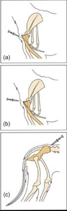
Figure 19.6 Sites for marrow aspiration include proximal humerus (a); manubrium of sternum (b); and iliac crest (c).
Obtaining good quality marrow samples requires sterile procedure and appropriate equipment. The skin is aseptically prepared, local anesthetic is infiltrated into the skin, subcutaneous tissue, and periosteum, and a small skin incision is made. Sternal aspirates can be collected with a regular 20-gauge hypodermic needle due to the thin cortex of this bone.
This can be very helpful in critically ill patients not suited for general anesthesia, or in obese patients where other sites are difficult to precisely locate. Penetrating the sternal marrow cavity with a hypodermic needle requires much less force than penetration of the cortex of pelvic, humeral, or femoral bone. Typically, only 0.1 to 0.3 mL of sternal marrow can be aspirated but that is sufficient for preparing multiple slides. To aspirate marrow from other sites, special needles of hardened steel (e.g. Rosenthal needles) with a stylet that occludes the lumen during bone penetration are required. Such needles must be sharp, the stylet must be precisely aligned with the shaft of the needle, and considerable force is needed to gradually advance the needle with forth-and-back rotation through the bone cortex. Single-use needles are popular because they are consistently sharp and do not require re-sterilization, but their use is more costly. Multi-use needles can be sharpened and autoclaved for multiple aspirations. Once the needle is seated firmly in bone, the stylet is removed, and a 6 or 12 mL syringe containing a few drops of 1% ethylenediaminetetraacetic acid (EDTA) is tightly attached. Vigorous aspiration with the syringe creates negative pressure that disrupts sinusoids and dislodges cells in the marrow cavity to yield gradually appearing, thick bloody fluid in the syringe. Generally, 0.2 to 0.5 mL of marrow in the syringe is sufficient for at least 5 smears, and the remaining fluid may be placed in EDTA-containing tubes for automated hematology analysis, flow cytometry, and/or PCR. Aspirating greater volumes of marrow induces progressively more hemorrhage from disrupted sinusoids, and is not recommended due to dilution of marrow cells. Marrow can be aspirated without EDTA in syringes if the operator is experienced, the samples are obtained quickly, and slides are prepared rapidly before the sample clots.For core biopsy, a longer and wider needle suitable for cutting a core of marrow containing trabecular bone with interspersed hematopoietic and adipose tissue is required (e.g.
11-, 13-, or 15-gauge Jamshidi needle). Marrow core biopsy should be performed prior to or at a different site than marrow aspiration to avoid areas with hematopoietic architecture disrupted from aspiration. The skin at the chosen site is aseptically prepared and infiltrated with local anesthetic as for aspiration, a stab incision is made, and the core biopsy needle with stylet in place is ‘drilled’ into the cortex using steady pressure with forth-and-back rotation at approximately a right angle to the site of bone penetration. Once the needle is seated firmly in the cortical bone, the stylet is removed, and the ‘empty’ needle is advanced for 2 to 3 cm to cut out a cylindrical core of marrow tissue. Penetration of the distant cortical bone by the needle without the stylet in place is very difficult; therefore, there is little danger from advancing the empty needle for 3 to 4 cm in the marrow cavity of a medium-size dog. Next, it is attempted to break off the distal part of the cylindrical core by rotation and slight lateral movements of the needle. Once it is thought that a core has been captured in the lumen, the needle is gradually retracted from the bone. The core itself is recovered from the lumen of the needle by gentle retrograde propulsion with a blunt wire, which is supplied with the core biopsy needle. The needle itself is tapered toward the tip to facilitate retention of the core; therefore, pushing the core out forward would crush the tissue. Further details on marrow collection techniques are available elsewhere (Bienzle, 2012, Abrams-Ogg et al., 2012, Defarges et al., 2013).