Necropsy: The Postmortem Examination
OBJECTIVES
• know the difference between an autopsy, necropsy, antemortem, and postmortem examination
• understand the reason a veterinary technician should be able to perform a necropsy
• know the safety precautions necessary for the protection of all personnel involved in a postmortem examination in all circumstances, whether it is a routine necropsy or one that may contain an infectious agent and has the potential to spread to animal and/or human populations
• understand the record keeping requirements and methods utilized in antemortem and postmortem exams
• know what to look for, tests to be performed, and recordings necessary when doing an antemortem examination or when examining the carcass prior to starting the necropsy portion of the postmortem examination
• know how to perform a necropsy
• be able to section a tumor and know the fixation process prior to submission for histopathological analysis
• understand the basic tenets of forensics and how it applies to the postmortem examination in animals
MATERIALS
• deceased animal, not preserved
• Mayo dissecting scissors
• serrated postmortem scissors or other serrated scissors
• probe
• 1 ? 2 thumb forceps or Adson tissue forceps
• #3 or #4 scalpel handle and blades
• bone-cutting forceps
• rubber gloves
• glass jars
• 10% formalin solution
• glass slides
Introduction
The purpose of a necropsy is to determine the cause of death of a patient.
In some cases the antemortem information gained through the physical exam, laboratory results, and other diagnostic procedures will provide sufficient knowledge to know what exactly caused the patient's death. In these situations, observations of pathological changes in the body obtained during a postmortem exam will help you ascertain what else may have contributed to the death, and the extent of the lesions involved. In other situations you may have virtually no knowledge of the patient prior to its death, and the necropsy is expected to solve the mystery of the patient's demise.The necropsy procedure outlined by the AVMA-CVTEA guidelines for accreditation is part of what every veterinary technology student must learn. The purpose of this knowledge is to
340
enable a technician to perform a necropsy by opening the body cavities; then the veterinarian or pathologist can view the organs in situ and lay them out on a tray after removal from the body so further examination is possible. At this time a diagnosis can be made and/or lesions noted that must be sectioned and placed in formalin to be sent to a laboratory for histopathological analysis.
Technicians may also be part of a forensics team that goes beyond just determining the cause of death of the animal, but also tries to recreate the events that occurred close to death, and to reveal any part that trauma, poison, disease, or neglect may have played. This reconstruction of events must fall within legal and possibly medical-criminal guidelines.
Necropsy Considerations
Safety should always be the primary concern. The potential always exists for three types of hazards: biological, chemical, and physical. Depending on the situation—whether the necropsy occurs in a veterinary hospital, involves a farmer’s animal in the field, or the investigation of a death of unknown cause of a wild bird or on a poultry facility, appropriate clothing is required and can vary from a protective smock or apron and wearing rubber gloves, to the highest-level, fully encapsulated suit with a selfcontained breathing apparatus.
The nature of the circumstances surrounding the animal’s death, what diagnostic procedures have been done antemortem, and knowledge about the patient will dictate the extent of the workup prior to the necropsy procedure. The best time to perform a necropsy is as soon after death as possible. Even with refrigeration, lesions become more and more difficult to assess as the postmortem period lengthens.
The animal’s body should never be frozen, and neither should the specimens obtained during a necropsy.Good record keeping is essential, whether you are using standard medical records or specific necropsy forms. If not previously recorded on medical records, the signalment information should be documented; this includes the owner’s name and address, the animal’s name or number, species, gender, age, breed, color, markings, and other pertinent information that would specifically identify the animal. The initial part of the physical postmortem exam documents the animal’s measurements, including length: tip of the nose to base of the tail; height: to top of the shoulder; and girth depending on the species. Record information from tags, bands, and tattoos. Scan the carcass for microchips. Describe all abnormalities observed on the animal’s body and collect samples where possible. Note that any interpretation of lesions should not appear in the description. Make an assessment of the animal’s body condition; was the animal of normal weight, underweight, cachexic, overweight, or obese? Also note the state of muscle rigor present.
Ideally the body should be weighed and its temperature recorded. If you are taking field notes, record the surface the animal was lying on and the condition of the ground around the animal. In addition, note the air temperature. Organ weight data can be recorded when collected during the necropsy.
Radiographs of the body are optional and should be done if this is a forensic investigation. Items to note are newly broken or dislocated bones, and healing and healed fractures. Look for the presence of foreign objects. Note any patterns of possible abuse. Making notes on dental development, bone calcification, ossification centers, and epiphyseal closure can provide accurate age information, especially in pre-adults.
The surface of the body should be observed for scars, abnormalities, discharges, discolorations, nodules, masses, and abnormalities.
Comb the animal and hold a piece of paper under the area you are combing to collect and observe the debris obtained. Shave, count, and measure all wounds and lesions. Bruising and bleeding in the tissues near wounds indicate that they occurred before the animal died. Look for, collect, and preserve any external parasites. Note the conditions of and any changes in the eyes, ears, nose, mouth, anus, urinary and genital tracts, and other openings.In a forensic investigation, photographing the body and the surrounding area is necessary. Include any significant findings and all lesions, both on the surface of the animal and those observed during the necropsy procedure. Placing a ruler adjacent to a lesion when photographing it will document its size.
Collect representative samples as indicated, including such things as blood, insects, mites, bone marrow, gastrointestinal contents, feces, and urine. You can take bone marrow from the femur, rib, sternum, or vertebrae. Blood may be collected directly from the heart or major vessels, and urine from the urinary bladder. These samples can be helpful because certain laboratory values remain stable after death. You should save gastrointestinal contents for identification and to screen for toxic substances. During the necropsy, normal saline should be used to rinse organ surfaces, as water will cause artifacts. Take microbiological samples as soon as possible during the necropsy by using sterile instruments to cut away contaminated surfaces; then collect specimens with additional sterile instruments or culture swabs. Take samples from any abnormal areas. Even though autolysis can cause artifacts to occur in tissues, it is better to obtain a tissue sample and let the pathologist decide if the lesions noted were a result of a disease process or due to postmortem changes.
EXERCISE 17.1 THE NECROPSY
The procedure demonstrated as depicted in the photos was performed on a cat euthanized at the owner’s request. The presumptive diagnosis, which was confirmed on necropsy, was hepatic lipidosis.
This cat was used to show the organs in a more natural state as compared to the preserved cats used for dissection. The necropsy procedure described here is a standard method that can be used on any species or size of animal. A review of the literature on this procedure revealed only slight variations in technique and order from the procedure listed here. It is written as though a graduate veterinary technician is performing the necropsy, with a veterinarian or pathologist in attendance as the supervisor and diagnostician.Procedure
1. Begin the procedure using sharp, clean necropsy instruments such as those pictured in Figure 17.1. If microbiological samples need to be taken, have a set of sterile instruments and culturettes available. Be sure to have all other necessary supplies and specimen containers on hand and readily available, including jars of formalin. As previously mentioned, if applicable, have someone take notes for you, or speak into a recording device for later transcription. Observe and note any lesions on the surface of the body and on any orifice. In Figure 17.2 note the jaundice (yellow color) of the skin together with petechial hemorrhages.
2. Placing the animal in dorsal recumbency, make a midline incision from the tip of thejaw to just below the anus. If an abdominal infection is suspected, use sterile instruments to prep the outside of the skin, make a small incision, and then introduce a sterile culturette into the abdomen to obtain an uncontaminated microbiological sample.
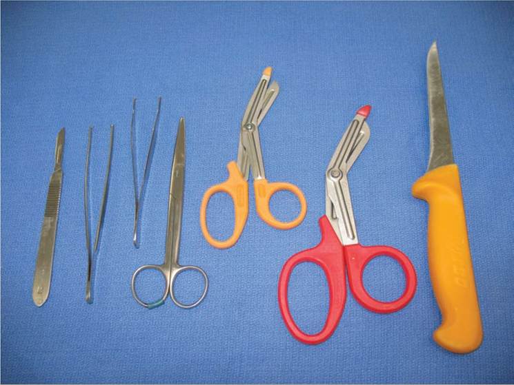
FIGURE 17.1: Instruments needed for the necropsy procedure.
3. Using scissors or a scalpel, dissect and spread the skin from the abdomen, thorax, and neck area, exposing the underlying tissues and lymph nodes. Note the amount and color of the subcutaneous adipose tissue; in this cat the fat is icteric (jaundiced) as a result of bile pigment infiltration (Figure 17.3).
4. Carefully cut the muscles between the scapula and the thorax, and the muscles and soft tissue surrounding the hip joints, to allow the pelvic and thoracic limbs to lie flat against the table, thereby stabilizing the cat in dorsal recumbency. Ideally, do not cut any vessels or nerves within the front or hind limbs (Figure 17.4).
5. In the neck area, remove the muscles ventral to the trachea, and examine the thyroid and parathyroid glands on both sides (Figure 17.5). Then locate and examine the salivary glands and regional lymph nodes.
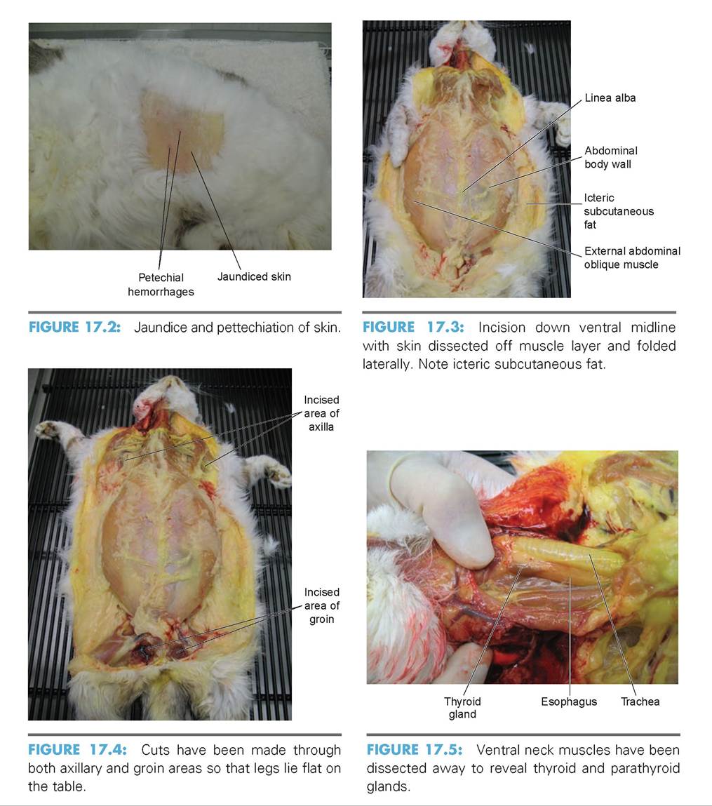
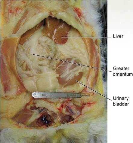
FIGURE 17.6: Abdomen has been opened to reveal the abdominal organs in situ. Note the centimeter marks on the scalpel handle.
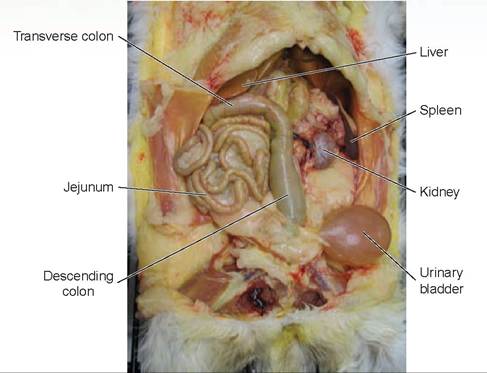
FIGURE 17.7: Greater omentum has been removed and organs are being explored within abdominal cavity.
6. To completely expose the abdominal contents, use your scissors to make a cut through the abdominal wall from the ventral midline incision, caudolaterally on both sides adjacent to the last ribs, to about midway on the lateral sides. At the cranial border of the pubis, make a similar cut from the midline incision laterally on both sides, and then fold the body wall out of the way to view the abdominal organs in situ (Figure 17.6). Note that the use of a scalpel handle marked in centimeters can be used to identify the relative sizes of the various organs.
7. Excise or reflect the greater omentum so that the abdominal organs can be observed. At this point, the veterinarian or pathologist will want to observe the organs (Figure 17.7). Methodically examine all of the viscera in situ, and observe their anatomical positions and sizes. Record any abnormalities noted. Observe the fluid in the abdomen. (The lateral cuts previously described are only made to the mid-lateral abdomen so that the fluid does not flow out.) Record any abnormalities in fluid quantity, color, consistency, or odor. Samples of this fluid can be taken for cellular content and other laboratory tests. Locate and examine both adrenal glands (Figure 17.8). If wound paths are present, use rods to demonstrate them, and then take photographs. In this cat, note the color of the liver. It should be dark reddish-brown; instead, it is a speckled yellow and red color, indicating that fatty infiltration has occurred in the parenchyma between the hepatocytes.
8. Verify the existence of negative pressure within the thoracic cavity by making a small stab incision through the diaphragm and listening for the rush of air into the pleural space. Ideally, this procedure should be done through the non-muscular portion of the diaphragm; any fluid present in the pleural cavity would drain out. In this cat, the puncture was done more ventrally because fluid was known to be present (Figure 17.9). At this time the puncture hole can be prepped and microbiological samples obtained from the thoracic cavity.
9. Using bone cutters (or, on smaller animals, serrated scissors), remove the ventral one-third of the rib cage (Figure 17.10). Observe the thoracic viscera in situ for correct anatomical position and size. If any abnormal fluid is present, record its quantity, color, consistency, and odor. Samples of that fluid can be taken and sent to the lab for cellular examination and other laboratory tests as required. In this cat, note the presence of a large quantity of icteric pleural effusion (fluid that has leaked out of blood vessels and lymphatics into tissues or a body cavity). Again, demonstrate wound paths, as described for the abdomen.
If the owner wants to take the animal home for burial, perform a cosmetic necropsy. Instead of removing the ventral one-third of the rib cage, make an incision either through the sternum or
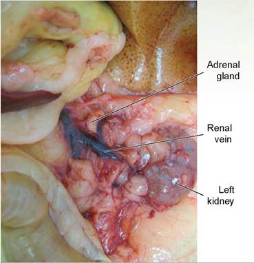
FIGURE 17.8: Left adrenal gland is examined. FIGURE 17.9: Incision is being made through the diaphragm.
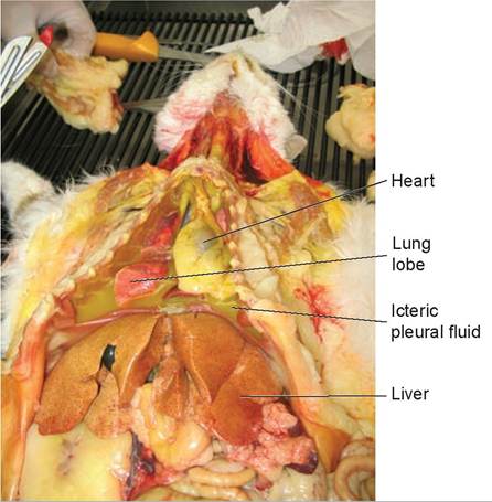
FIGURE 17.10: Ventral one-third of the chest wall has been removed. Note the icteric pleural effusion in the thoracic cavity.
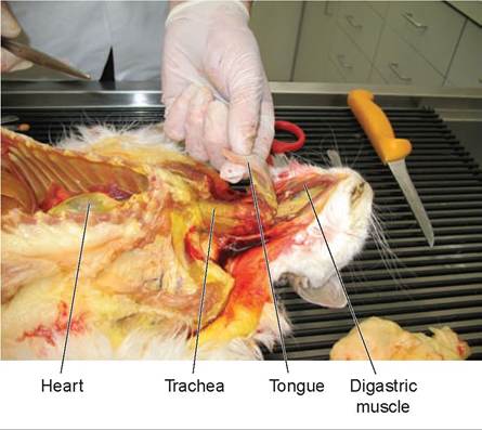
FIGURE 17.11: The tongue has been removed from the oral cavity and the dissection for removal of the viscera of the neck and thoracic cavity is being initiated.
adjacent to the sternum; then hold the chest cavity open with self-retaining retractors. After your examination of the thoracic and abdominal viscera is complete, sew the body walls and skin back together again, and then send the animal home for burial.
10. Dissect through the soft tissue adjacent to the digastric muscles along the mandible, through to the mouth and around the tongue and larynx. Pull the tongue down through the incision area; then remove the tongue, larynx, trachea, esophagus, lungs, and heart as a unit, tying off the distal esophagus to prevent gastric spillage (Figure 17.11). Examine the oral cavity, teeth, pharynx, and tonsils.
11. Lay the removed organs on a surface for examination. Incise the pericardial sac and observe for abnormal fluid within; then reflect it over the base of the heart (Figure 17.12). Next, examine the following: the base and apex of the heart, coronary vessels, great vessels, atria, and ventricles. Dissect the heart along the path of blood flow, similar to the way it was done during the dissection of the sheep’s heart. Remove any blood and examine the endocardium, the thickness of the heart walls, and the valves, and look for abnormalities that would indicate problems with blood flow, such as a ventricular septal defect (Figure 17.13). The heart of this cat does not appear enlarged, although samples of it should be sent in for histopathological examination to rule out hypertrophic cardiomyopathy.
12. Locate and section the thymus and the lymph nodes of the cranial thoracic viscera (Figure 17.14). Examine the external surface of the trachea, and open it from the larynx to the main bronchi (Figure 17.15). Palpate all lung lobes, and dissect through the lobar bronchi. Next, examine and open the esophagus (Figure 17.16). In this cat, there are red areas in the lungs indicating inflammation and consolidation, and the dorsal aspect of the intermediate lobe has an unusual lesion.
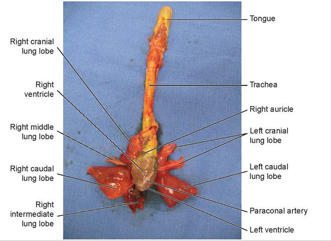
FIGURE 17.12: The viscera, including the tongue, esophagus, larynx, trachea, lungs, heart, and pericardium, have been removed from the mouth, neck, and thorax. The pericardium in this photo has already been removed.
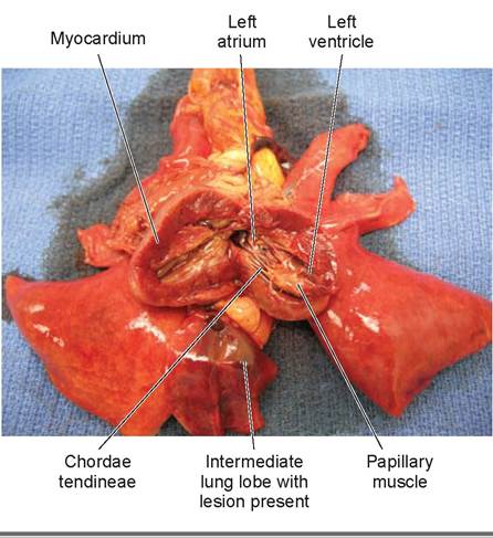
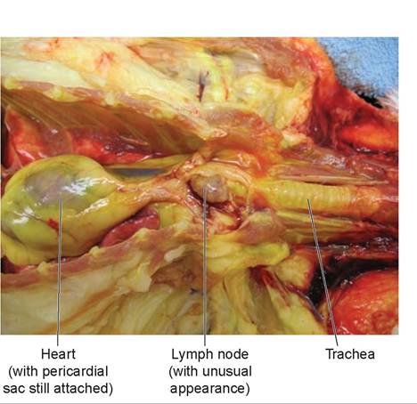
FIGURE 17.14: Photo showing an enlarged and unusual lymph node in the cranial mediastinal area. Head of the cat is toward the right.
FIGURE 17.13: Photo of an open left atrium and ventricle (the right atrium and ventricle has already been opened).
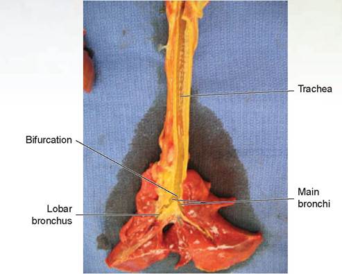
FIGURE 17.15: Incised trachea and bronchi extending into the lung lobes.
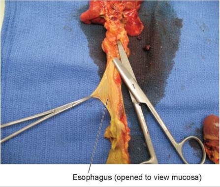
FIGURE 17.16: Using scissors to open the esophagus attached to the dorsal aspect of the trachea.
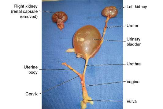
FIGURE 17.17: Dissected urogenital system from the vulva to the kidneys. This cat was spayed so the vagina, cervix, and part of the uterine body were present.
13. Using bone cutters, locate by palpation each obturator foramen and cut the pubis cranially and caudally on both sides, opening the pelvic canal. Using a scalpel, incise the skin and subcutaneous tissue around the external genitalia. Free both the kidneys and ureters from their attachment sites. Bluntly dissect the external genital tract, urethra, and, in the female, dissect the vagina, cervix, and body of the uterus (include the horns and ovaries if still present) off of their attachment to the rectum. In the male, remove both the testes and spermatic cord from their attachments at the proximal prostate area. Remove the kidneys, ureters, bladder, urethra, and genitalia, and place them on a clean surface for examination. Reflect the renal capsule from each kidney, and examine the ureters and renal vasculature. Cut one kidney longitudinally, and the other transversely; this was not done in Figure 17.17, so that it could show the entire genito-urinary tract. Then extend each incision into the renal pelvis and ureter. Then serially section each kidney and ureter. Open the bladder, urethra, and prostate gland (if a male). Then examine the genitalia.
14. Examine the anus and anal glands; then, using a scalpel, incise the surrounding skin and subcutaneous tissues. Remove the entire abdominal viscera as a unit. Then examine the remaining lymph nodes and the abdominal aorta.
15. Lay the organs on a clean surface. Next, examine the entire pancreas. Then open the distal esophagus and stomach, and examine the contents and lining (Figure 17.18). Collect samples of the contents, as necessary. Incise through the pylorus to the area of the hepatopancreatic ampulla. Apply pressure to the gallbladder while observing for bile expulsion through the major duodenal papilla (Figure 17.19). Open and examine the gallbladder. Then open and examine the
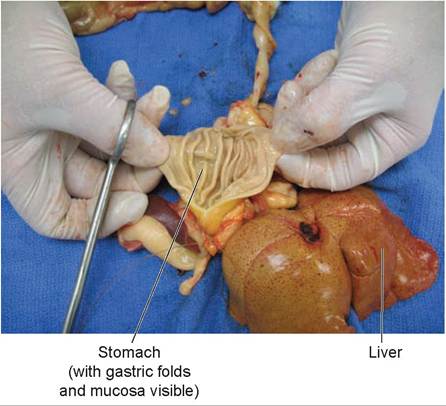
FIGURE 17.18: The gastrointestinal tract has been removed. The dissection starts with the opening of the stomach. Normal contents and gastric folds were noted.
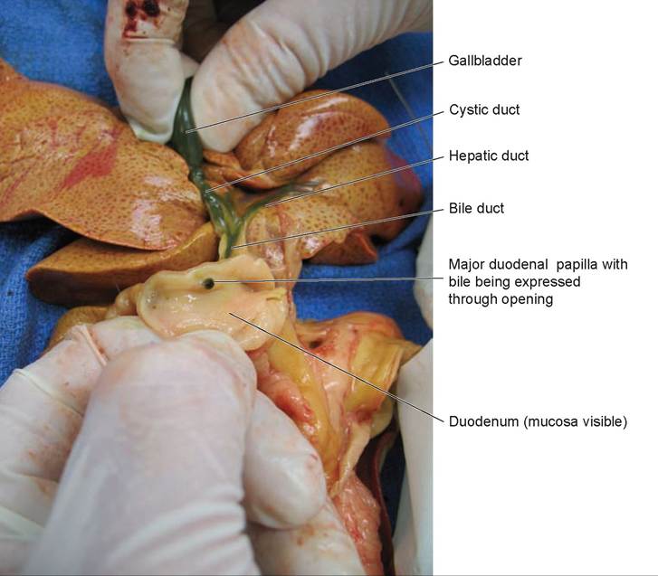
FIGURE 17.19: The duodenum has been opened and the major duodenal papilla with green bile coming through the opening is demonstrated after compressing the gallbladder. hepatic artery and veins, and the portal vein (visceral surfaces). Serially section the liver and spleen (Figure 17.20). Samples can be taken in areas containing lesions. Examine the omentum, mesentery, root of the mesentery, and mesenteric lymph nodes. Open the duodenum and continue through both the small intestine and large intestine. In Figure 17.21, only a small part of the jejunum has been opened. Note that the mucosa is normal and lacks petechiation. Collect fecal samples as indicated.
16. Examine the hip, stifle, shoulder, and elbow joints for ruptured tendons and ligaments; then open them to look for arthritic changes and abnormal fluid (Figure 17.22). Document any evidence of trauma observed. For any bony swellings, the muscles should be stripped off and a section of diseased bone sent to the lab.
17. If there is any suggestion of neurological disease, the brain and spinal cord should be opened. In this exercise, the rest of the examination is optional. First, carefully remove the eyes intact. Then
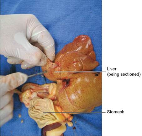
FIGURE 17.20: The liver is being sectioned. Note the abnormal light yellow speckled appearance of the liver tissue characteristic of fatty infiltration.
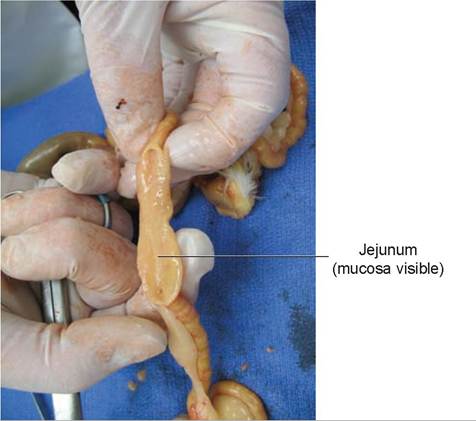
FIGURE 17.21: The jejunum is being opened along its length. Normal mucosa is visible.
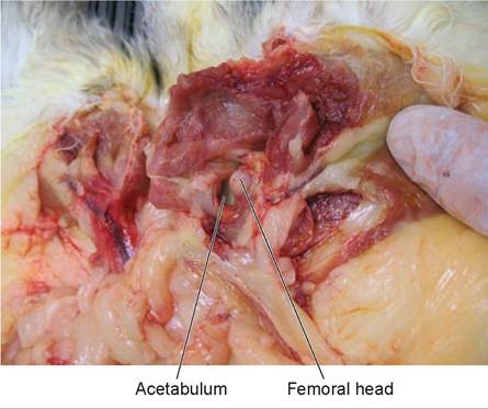
FIGURE 17.22: The coxofemoral joint has been opened and disarticulated with the femoral head and acetabulum visible. The pelvis is toward the left and the leg is toward the right.
remove the head. Make a dorsal incision from the nose to the foramen magnum on the midline, and reflect the skin ventrally. Next, transect and examine the ear canals. After excision of the temporal muscles, make lateral cuts from the foramen magnum cranially on each side, until they meet. Pry off the top of the skull and examine the internal dorsal surface. Turn the head over and let the brain fall gently into your hand or onto a surface, while carefully cutting the cranial nerves to free the brain from the skull. Then serially section the brain.
18. Saw transversely through the frontal and maxillary bones in front of the orbits to examine the nasal cavities and sinuses.
19. Return to the body cavities; examine the ventral aspects of the vertebrae. Remove the rest of the skin and observe the dorsal body musculature for any abnormalities.
20. In cases of spinal disease or trauma, cutting through the laminae and removing the dorsal spinal processes using a saw or bone cutter will open the spinal canal and allow removal of the spinal cord in one piece.
Hepatic lipidosis is a secondary disease caused when a cat stops eating (becomes anorexic). It is more likely tooccur in heavy or obese cats. Fatty infi ltration causes damage to hepatocytes and, ultimately, liver failure. Normalhemoglobin degradation cannot occur, and bile pigments back up into tissues, causing icterus in organs (and fl uids) and the jaundiced appearance of the skin. The necropsy was to determine the reason for the anorexia. The reason for the inciting cause was not readily apparent, and histopathological examination was needed. It is possible that some initial liver problems might have caused additional liver problems. Specimens taken for histopathological examination would have included the liver, the kidney, the entire intermediate lung lobe, the abnormal cranial thoracic lymph node, and a distal section of the right humerus, which was either very arthritic or contained an osteo- sarcoma.Unfortunately,the owner did not elect to have histopathology done.
EXERCISE 17.2 SECTIONING METHOD FOR TUMORS
Tissue samples obtained for lab analysis during the necropsy procedure must be prepared in such a manner to ensure that the fixative solution (10% formalin) penetrates sufficiently deep to prevent autolysis in the center of the specimen. Formalin can be expected to penetrate only about 1 cm in most tissues. For a tumor found during a necropsy or removed during surgery, it is often necessary to preserve the entire structure to identify its borders and verify that complete excision occurred. The following exercise will demonstrate the proper method of tissue preparation to ensure adequate preservation throughout the specimen.
1. Using a crabapple, make serial slices that yield sections no greater than 1 cm thick. Do not cut all the way through the crabapple. Instead, leave a 1-cm thick area at the bottom of the cuts so that the fruit remains in one piece (Figure 17-23). If this were a tumor, a pathologist would be able to view the tissue in its entirety in its proper structural alignment, and to observe all its borders, and the formalin would be able to penetrate the entire tumor.
2. When fixing a piece of tissue, the amount of fixative needed is 10 times the volume of the tissue. Accordingly, a 1 cubic centimeter (cc) piece of tissue must be placed in 10 milliliters (ml) of fixative. To find a tumor’s volume in cubic centimeters, a technician could measure it and calculate it, or weigh it and utilize the following estimate: assume 1 gram (g) of tissue displaces 1 cc of fluid, therefore a tumor weighing 10 grams could be estimated to have a volume of 10 cc. Weigh your crabapple in grams and fill in the blanks below to estimate the amount of fixative you would need.
a. weight of crabapple:_____________ g =_____________ cc
b. _________ cc ? 10 =____________________ ml of fixative required.
c. What is the minimum number of slices necessary for a tumor that is 8 cm long?
Answer:_________________________________________________________________________
d. How many milliliters of fixative are necessary for a perfectly round tumor that is 4 cm in diameter? The equation for the volume of a sphere is V=4∕3πr3, where V= volume, r=radius.
Answer:_________________________________________________________________________
3. The tissue is fixed for 24 hours, and then it can be transferred to a small container filled with fixative for shipping. The answer to question c is seven slices, and the answer to question d is 335 ml.
Note that in “d” above, for simplicity’s sake, we used a perfectly round sphere. If the tumor were an ellipsoid, or egg-shaped, the formula would have been V=4∕3πr1r2r3 (see figure 17.24). It is obvious now thatjust weighing a tumor or mass and using the approximation of converting the weight directly to cc’s is much easier than doing these calculations.
There may be a time when the organ or tumor is too large and/or you do not have enough formalin to ensure adequate preservation. In this case, placing the specimen in a bag filled with physiologic saline solution (and then double bagging it) will suffice if you can get it to a lab as soon as possible.
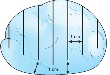
FIGURE 17.23: Method of sectioning of a tumor for fixation.
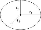
FIGURE 17.24: The three radii used to calculate the volume of a elliptoid-shaped object.
Clinical Significance
Forensics is an area of veterinary medicine in which all veterinarian must be knowledgeable especially considering the possibility of bioterrorism or zoonotic disease outbreaks. According to Dr. Lonnie King, Acting Director of the National Center for Zoonotic, Vector-Borne, and Enteric Diseases, infectious organisms cause some 1,620 human diseases, 60 percent of which are zoonotic. In addition, of the 35 major human disease outbreaks over the past two-and-a-half decades, 75 percent were zoonotic. Most states have procedures in place or are developing procedures to address disasters, terrorism, and zoonotic diseases. Emergency response teams combining veterinarians and veterinary technicians respond to the plights of animals caught in deadly situations and to emergent strains of any zoonotic diseases that might enter their state or area. Training in proper procedures, in the use of protective equipment, and in the team’s activation and response are of critical importance.
Safety is again the prime concern. If something killed an animal, the aim is to prevent it from killing again. The common procedure is to institute a three-layered border to isolate and secure a scene, whether it is a crime scene or the scene at which an animal or bird is found dead of unknown causes. The outer border is the safe area for bystanders, media, and nonessential personnel; the middle zone is a buffer and work area; and the inner zone is the core area or the site where the event occurred. Barricades consisting of tape or ropes must be deployed to prevent unauthorized people from entering the area and potentially contaminating the site or themselves. A cleansing area for disinfecting gear and removing dangerous materials must be set up. Levels of protection required vary with the nature of the event, and generally too much protection is better than not enough. Disposable coveralls, gowns, boot and shoe covers, and facemasks are best, but using a set of gear that is dedicated for this job and then disinfected is also acceptable. Even using a disposable pen to write with may prevent accidentally taking dangerous disease agents home or back to the office.
Experience will tell us when “something isn’t right.” This may be especially true with odors, as many conditions have a characteristic odor. Always treat unrecognized odors as though you are dealing with a toxic substance. Assume the animal has an infectious disease until proven otherwise.
If a disease is suspected that has great risk of environmental contamination, either a diagnosis must be obtained prior to the necropsy or it must be performed under strict quarantine conditions. Anthrax is an example of a bioterrorism weapon. Opening a carcass with anthrax will induce the bacteria to form spores that are then dispersed throughout the environment. If anthrax is suspected, get blood smears first from peripheral areas or from swellings, and take them to a lab for examination prior to opening the carcass during a necropsy. If the diagnosis is confirmed, the carcass must be quarantined and destroyed properly, such as incineration.
Determination of time of death is dependent on body temperature, which in turn depends on ambient environmental temperature. Body weight and an animal’s fat layer all factor into body temperature change. Initially, body temperature drops; however, after the onset of putrefaction due to metabolic activity of bacteria and other decomposing organisms, the body’s temperature will rise again. Rigor mortis depends on the temperature and concentrations of lactic acid. High metabolic activity just prior to death (e.g., running) leads to higher metabolic levels of lactic acid and thus shortens the time required for muscle rigor to develop. Higher environmental temperatures also shorten this time. In temperate regions, the following is a general rule of thumb that may help in estimating the time of death (but use it with caution): Warm means dead for not more than eight hours; stiff means dead for not more than three days.

---------------- his final story is almost embarrassing to tell. When I was between my third and fourth years X of veterinary school I worked as a technician in a local veterinary hospital in Ft. Collins, (j∕ Colorado, where we encountered the case of a dog with severe dyspnea. The owner told us that he had brought the dog in as soon as he noticed its condition. A thoracic radiographic series showed multiple radio-opaque masses scattered throughout the lung field. It appeared to be either primary or metastatic neoplasia, and the owner decided not to biopsy any of the thoracic masses and instead elected euthanasia. The veterinarian in charge of the case recommended that we send the dog to the diagnostic lab at Colorado State University. I made the arrangements and took the body to the pathology lab.
Upon opening the chest, we found granulomatous lesions characteristic of a deep fungal infection, which the lab confirmed to be cryptococcosis, caused by the organism Cryptococcus neoformans. It was not surprising this dog was dyspneic; very little normal lung tissue remained. Furthermore, the owner must not have been paying much attention to this dog because this disease does not get to this level overnight.
About a week or so later, I noticed a red, pustular lesion on the front of my right forearm. Of course, I immediately got out my pathology book and read that cryptococcosis has multiple forms: respiratory, cutaneous, ophthalmic, and nervous. The fungus responsible is transmitted by inhalation of the yeast-like form of the organism, and in cats it usually colonizes the nose and sinuses before spreading to other organs. In dogs it has a predilection for the CNS and the eye but disseminates to more organs than it does in the cat. I discovered that no documentation exists that the spherical organisms found in tissues are directly transmissible from animals to humans. In other words, humans can get cryptococcosis, but only from the yeast-like soil-borne state. I was safe. At least, I thought so—until I went to a dermatologist.
“It's theoretically possible you got infected if you splashed cells from the dog's chest onto an already-open sore,” he told me.
“I didn't splash any tissue onto my arm,” I told him. He was not convinced. So much for applying scientific evidence and logic.
“Oh boy, this could be my first case of cryptococcosis,” he declared excitedly. “We'll have to do a punch biopsy.” He actually said this! I don't know where he went to medical school and learned his bedside manner, but I figured it had to be Lower Moronovia Medical School for the Terminally Insensitive.
Still, no matter how much I was sure he was full of dog-poo, in the back of my mind the “What ifs” kept creeping in. As it turned out, the biopsy came back negative; it was nothing more than a little bacterial infection. A course of antibiotics and I was fine.
As I think back on this, I was lucky, for “a little bacterial infection” could have been more than this. One of my veterinary school classmates developed a very resistant staph infection on his neck. As I remember, it took months and the use of an autogenous vaccine to get rid of it.
Early in my career I used to be more cavalier in my dealings with cat abscesses and the like, but now I wear protective gloves when dealing with all infections, and a mask and glasses for dentals. I am conscientious about wearing my protective gear during radiographic exposures, and about making sure that no part of me is in the direct beam, even when wearing gloves. Safety first, in all things!
Summary
In this chapter you have learned about the necropsy procedure: why it is necessary for veterinary technicians to know how to perform one, and how you might assist your veterinarian and add another skill to your repertoire. Safety has been stressed a number of times, both in terms of your personal safety but also that of the environment. You learned about antemortem examinations of the body and all they entail, plus the procedures involved in postmortem examinations. When it is necessary to send an organ or pathological specimen such as a tumor to a lab, you learned how to secion it.
review questions
1. What is the purpose of a necropsy?
2. Differentiate between antemortem and postmortem.
3. Why would a veterinary technician need to know how to perform a necropsy?
4. What is the primary concern in all postmortem exams?
5. Name the three types of hazards that might be encountered during a necropsy.
6. When is the best time to perform a necropsy?
7. Define signalment.
8. What parts of an animal should be examined prior to starting to cut?
9. What liquid should be used to clean the surfaces of the body or of organs, and why?
10.Name all the equipment and supplies needed to perform the necropsy procedure.
11.Why is the abdomen opened as described in the necropsy procedure?
12.Why is a puncture through the diaphragm made prior to opening the thorax?
13.Why is only the ventral one-third of the chest wall removed?
14.What thickness of tissue can formalin be expected to penetrate?
15. To ensure that a tumor that is 10 cm long and 5 cm thick is properly fixed, how many cuts of what depth would be required?

Bacha, W. J., Jr., and L. M. Bacha, Color Atlas of Veterinary Histology. 2nd ed. Baltimore: Lippincott, 2000.
Boyd, J. S., Color Atlas of Clinical Anatomy of the Dog and Cat. 2nd ed. Philadelphia: Mosby, 1999.
Cochran, P. E., Guide to Veterinary Medical Terminology. St. Louis: Mosby, 1991.
Coles, E. H., Veterinary Clinical Pathology. 4th ed. Philadelphia: W. B. Saunders, 1986.
Colville, T., and J. Bassert, Clinical Anatomy & Physiology for Veterinary Technicians. St. Louis: Mosby, 2002.
Cooper, E. L. and L. D. Burton, Agriscience Fundamentals & Applications. 3rd ed. New York: Delmar Thomson Learning, 2002.
Edwards, N. J., ECG Manual for the Veterinary Technician. Philadelphia: W. B. Saunders, 1993.
Evans, E. E., Miller’s Anatomy of the Dog. 3rd ed. Philadelphia: W. B. Saunders, 1993.
Evans, H. E., and A. deLattunta, Miller’s Guide to the Dissection of the Dog. 3rd ed. Philadelphia: W. B. Saunders, 1993.
Ettinger, S. J., Textbook of Veterinary Internal Medicine, Diseases of the Dog and Cat. Philadelphia: W. B. Saunders, 1975.
Frandson, R. D., and T L. Spurgeon, Anatomy and Physiology of Farm Animals. 5th ed. Philadelphia: Lea & Febiger, 1992.
Gilbert, S. G., Pictorial Anatomy of the Cat. 7th ed. Seattle: University of Washington, 1987.
Holmstrom, Steven E., Veterinary Dentistry for the Technician & Office Staff. 1st ed. Philadelphia: W. B. Saunders, 2000.
Hudson, L. C., and W. P. Hamilton, Atlas of Feline Anatomy for Veler ioar iaos. Philadelphia: W. B. Saunders, 1993.
Jenkins, T W., Functional Mammalian Neuroanatomy. Philadelphia: Lea & Febiger, 1972.
Kealy, J. K., Diagnostic Radiology of the Dog and Cat. 2nd ed. Philadelphia: W. B. Saunders, 1987.
Marieb, E. N., Human Anatomy & Physiology Inhoratory Manual. 7th ed. San Francisco: Benjamin Cummings, 2002.
Miller L. and Zawistowski S., Shelter Medicine for Veterinarians and Staff: Veterinary Forensics, Ames, Iowa: Blackwell, 2004.
355
McDonald, L. E., and M. H. Pineda, Veterinary Endocrinology and Reproduction. 4th ed. Philadelphia: Lea & Febiger, 1989.
Neal, K. G., and B. H. Kalbus, Dissection Guidefor the Cat (and Selected Sheep Organs). Minneapolis: Burgess, 1971.
Patten, B. M., Foundations of Embryology. 2nd ed. New York: McGraw-Hill, 1964.
Pratt, P.W., Principles and Practice of Veterinary Technology. St. Louis: Mosby, 1998.
Reagan, W. J., T G. Sanders, and D. B. DeNicola, Veterinary Hematology Atlas of Common Domestic Species. Ames, Iowa: Iowa State University, 1998.
Rebar, A. H., Handbook of Veterinary Cytology. St. Louis: Ralston Purina, 1978.
Reece, W.O., Functional Anatomy and Ihysif)lf)gy of Domestic Animals. 3rd ed. Philadelphia: Lippincott Williams & Wilkins, 2005.
Sandman, K. M., and J. Hatari. “Cranial Cruciate Ligament Repair Techniques: Is One Best?” Veterinary Medicine96(11): 850-856, 2001.
Sisson, S., and J. D. Grossman, The Anatomy of the Domestic Animals. 5th ed. Philadelphia: W. B. Saunders, 1975.
Stashak, T S., Adam’s Lameness in Horses. 4th ed. Philadelphia: Lea & Febiger, 1985.
Synbiotics Corporation. ICG Status-Pro & ICG Status-LH. San Diego: Synbiotics Corporation, 1998.
Tilley, L. P., and F. W. K. Smith, The 5-Minute Veterinary Consult, Canine and Feline. Baltimore: Williams & Wilkins, 1997.
Tortora, G. J., and S. R. Grabowski, Principles of Anatomy and Physiology. 9th ed. New York: John Wiley & Sons, 2000.
A
Abdominal, defined, 3
Abdominal aorta, 228
Abdominal oblique muscle, 167
Abdominal wall muscles, 167
Abducens nerve, 318
Abduction, defined, 4, 5
Abductor cruris caudalis muscle, 172
Abomasum, 197, 199
Absolute cell count, 83
Absolute refractory period, 304
Accessory digestive organs, 181
Accessory head, 162
Accessory lobes, 243
Accessory pancreatic duct, 193-194
Accessory sex glands, 283
Acetylcholine, 311 Acetylcholinesterase, 320 Achilles tendon, 174
Acidophilic cell parts, 27
Acromial part, 159
Actin, 149, 152
Action of the muscle, 154-155
Action potential, 303
Active transport, 20-21
Adduction, defined, 4, 5
Adductor brevis, 172
Adductor longus, 172
Adductor magnus, 172
Adductor muscles, 172
Adductor pollicis longus muscle, 165-166
Adenohypophysis, 268
Adenosine diphosphate (ADP), 177
Adenosine triphosphate (ATP), 21, 25, 177
Adipocytes, 62
Adipose tissue connective tissue, 54 described, 62 function, 63 hypodermis or subcutis, 92 location, 62-63 pigmented skin, 96 skin, 86 slide, 63-64 subcutaneous, 98
Adjacent, defined, 2
Adrenal cortex, 269
Adrenal glands, 270-271, 273
Adrenal medulla, 271
Adrenalin, 271
Adrenocorticotropic hormone (ACTH), 269
Adventitia, 182
Aging a dog or cat, 183-184
Agonist muscle, 320
Agranulocytes, 70
Air capillaries, 248
Aldosterone, 259, 270
Alimentary tract, 181, 182
Allantoic membranes, 285
Allantoic sac, 285
Allantois, 285
All-or-none principle, 304
Alpha cells, 272
Alveolar duct, 245
Alveolar sac, 245
Alveoli, 41, 43, 245
Amnion, 285
Amnionic sac, 285
Amorphous ground substance, 66
Ampullae, 283
Amylase, 202-203
Anatomical terms, 3-4
Anconeus muscle, 162
Androgens, 271
Anestrus, 285, 292
Angiotensin, 259
Antagonist muscle, 320
Antebrachium, defined, 3
Ante-mortem, 341
Anterior, defined, 1
Anterior chambers of the eye, 335
Anterior pituitary, 268
Anterior-posterior view, 6
Antidiuretic hormone (ADH)
endocrine system, 268, 270
urinary system, 259, 263-264
Antigenicity, 20
Antlers, 100
Anucleated red blood cells, 70
Aorta, 210, 217
Aortic arch, 225
Aortic valve, 218
Apex of the heart, 211
Apical foramen, 185
Apical lobes, 243
Apical surface, 41
Apocrine glands, 94, 96
Appendicular, defined, 3
Appendicular skeleton, 109
Apple, cross-sectioning, 35-36
Aqueduct of Sylvius, 319
Aqueous humor, 335
Arachnoid, 314
Arbor vitae, 314, 319
Arcuate artery, 255, 262
Arcuate vein, 262
Areolar or loose connective tissue, 54, 56-57
Arrector pili muscle, 96
Arterial system, 208
Arteries, 219, 223, 224-229
Arthrology, 139-140
Articular projections, 110
Arytenoid cartilages, 240
Ascending colon, 195
Ascending loop, 258
ATP. See Adenosine triphosphate (ATP) Atrial systole, 209
Atrioventricular bundle, 230
Atrioventricular node, 230
Atrioventricular valves, 209 Auditory tubes, 188, 239, 327
Auscultate, 251
Autolysis, 24, 32
Autonomic nervous system, 299, 310-313
Autophagy, 24
Axial, defined, 3
Axial skeleton, 109
Axillary, defined, 3
Axillary artery, 227
Axillary vein, 223
Axon, 28, 81, 300, 302
Axon hillock, 82, 303
Axonal terminal, 301
Azurophilic granules, 74
Azygos vein, 221
B
Back
deep back muscles, 176-177
deep muscles, 161
superficial muscles, 158-159
Band neutrophils, 71, 74
Bars, 100
Basal cells, 49, 53
Basal lamina, 41
Basal surface, 41
Base of the heart, 211
Basement membrane
epidermis, 87
epithelial tissue, 41
simple columnar epithelium, 46
simple squamous epithelium, 42
stratified columnar epithelium, 52
stratified squamous epithelium, 49 urinary bladder or transitional epithelium, 53
Basihyoid bone, 240
Basophilic cell parts, 27
Basophils, 56, 70, 71, 75
Beta cells, 272
Biceps brachii muscle, 162
Biceps femoris muscle, 170
Bicornuate, 288
Bicuspid valve, 218
Bile, 182
Bile duct, 182, 195
Bipolar neurons, 301
Birds. See Fowl
Bitch, mammary gland, 104
Blood, 54, 70-76
Blood pressure, 219, 233-234 Blood-brain barrier, 322
Blood-vascular system, 207, 219
Body of the stomach, 190
Body of the uterus, 280, 288 Boideus thoracic muscles, 161
Bone marrow cavities and spaces, 109 Bones. See also Skeletal system basihyoid bone, 240
cancellous (spongy) bone, 109, 114-116 coffin bone, 100
compact bone, 109, 111-114 comparative osteology, 130-139 carpus, 134
chicken skeleton, 138-139 feet, 137
foreleg, 133
hind leg, 135
skulls, 130-132
tarsus, 136 connective tissue, 54, 76-77 distal sesamoid bone, 100 erosion of, 109 flat bones, 110 functions, 109 Haversian system of, 112 histology, 111-116
cancellous (spongy) bone, 114-116
compact bone, 111-114 irregular, 110 lacuna bone, 77 long bone parts, 110, 111 metacarpal bones, 125-126 nasal turbinate, 239 navicular bone, 100 pneumatic, 110 process, 110 projections, 110 remodeling, 109 short bones, 100, 110 types of, 110 woven bone, 109
Bony orbit, 332 Bowman’s capsule, 37, 43, 44, 258 Brachial, defined, 3
Brachial artery, 227 Brachial plexus, 305, 306 Brachial vein, 223
Brachialis muscle, 162 Brachiocephalic artery, 226 Brachiocephalic trunk, 226 Brachiocephalic veins, 222 Brachioradialis muscle, 165 Brain
anatomy, 314-319 brainstem, 313, 314 nerve cells, 82 nervous system, 299 physiology, 313-314
Branched cardiac muscle, 79, 80 Broad ligament, 281, 288 Bronchial sounds, 251
Brown fat, 62 Buccal, defined, 4 Bulbar conjunctiva, 332 Bulbourethral glands, 283, 292 Bulbs of the heel, 103 Bulbus glandis, 283
C
Calcium measurements, 142-143
Canal, 110
Canaliculi, 77, 113
Cancellous (spongy) bone, 109, 114-116 Capillaries, 45, 71
Capillary bed, 219
Carcinomas, 144
Cardia, 190
Cardiac lobes, 243
Cardiac muscle, 79-80, 147, 152-153
Cardiac region, 190
Cardiac sphincter, 190
Cardiovascular system
arteries, 224-229
blood pressure, 219, 233-234 blood-vascular system, 207 clinical significance, 234 electrocardiogram, 230-233 heart
anatomy, 211-218
cardiac cycle, 209-210 chambers, 209 function, 40, 207
overview, 207, 209-210
phases, 209
overview, 207-208
vascular system, 219
veins, 220-224
veterinary vignettes, 235-236
Carina, 242
Carotene, 96
Carpal pad, 97
Carpus, cat skeleton, 125-126
Cartilage connective tissue
blood, 70-76
bone, 76-77
elastic cartilage, 67-68 fibrous cartilage, 68-70 histology, 54 hyaline cartilage, 66-67 overview, 66
Cat
aging, 183-184
artery dissection, 224-229 blood, 71, 73
claw, 98
description using anatomical terms, 7-8 electrocardiogram, 231
Cat (continued )
endocrine gland dissection, 272-274 foot pads, 96-98
kidney dissection, 259-262 lower respiratory system, 242-247 mammary gland, 104 necropsy, 343-351 peripheral nerve dissection, 305-310 reproductive system dissection
female, 288-290
male, 290-292
skeletal musculature, 154-177 abdominal wall muscles, 167 chest, superficial muscles, 155-156 deep back muscles, 176-177 front leg muscles, 161-166 hind leg, deep muscles, 175 hind leg, superficial muscles, 167-171 neck, superficial muscles, 157-158 posterior leg muscles, 174 shoulder and back, deep muscles, 161 shoulder and back, superficial muscles, 158-159
thigh, deeper muscles, 172-174 skeleton, 116-129
carpus, 125-126 femur, 127-128 fibula, 128 humerus, 124 metacarpal bones, 125-126 metatarsals, 129 os coxae (pelvis), 126-127 phalanges, 125-126, 129 radius, 124-125 ribs and sternum, 119 scapula, 123-124 skull and mandible, 120-123 tarsus, 128 tibia, 128 ulna, 125 vertebrae, 116-119 topographical anatomy, 9 upper respiratory system, 239-242 vein dissection, 220-224
Cauda equina, 305 Caudad, defined, 2 Caudal, defined, 2, 3 Caudal artery, 229 Caudal colliculi, 314
Caudal lobes, 243
Caudal mediastinum, 242
Caudal mesenteric artery, 229
Caudal thyroid artery, 226
Caudal vein, 224
Caudal vena cava, 216
Caudally, defined, 2
Caudate lobe, 190
Cavities, 110
Cecum, 195
Celiac artery, 228
Cell
absolute cell count, 83 alpha, 272 anatomy, 20 anucleated red blood cells, 70 basal, 49, 53
beta, 272
cell body, 28, 300
cell junctions, 21
cell processes, 28
clinical significance, 29 columnar, 41 connective tissue, 54 cross-sectional appearance of a sheet of cells, 38
cuboidal, 41, 45 cut at angles, 35 cytoplasm, 20, 22 defined, 19 delta, 272
differential cell count, 70 embryonal mesenchymal, 109 endothelial, 42
erythrocytes (red blood cells), 70, 74 germ, 279 glial cells, 299 goblet cells, 48, 49
ICSH, 269 intercalated, 259 interstitial (cells of Leydig), 282 juxtaglomerular, 259
Kupffer cells, 182 Langerhans’ cell, 87 leukocytes (white blood cells), 70, 83 mast cells, 56, 57
Merkel cells, 87
mesenchymal cells, 55 mesothelial, 42 mucosal cells, 48-49 multinucleated, 21, 149 muscle cells, 79, 149 myoepithelial, 259 nerve cells, 81, 82 pancreatic islet, 272 parts of, 20-26
cilia, 26
cytoplasm, 20, 22 flagella, 26 identification using microscope, 27-29 inclusion bodies, 20, 26 nucleus, 20, 21-22
organelles, 20, 22-26 plasma membrane, 20-21
plasma cells, 56, 57 principal cells, 259 processed, 302 purpose of, 19 relative cell count, 83
Renshaw cell, 320
reticular, 65
Schwann cells, 304
sliced, 34
spindle-shaped, 81, 153 squamous, 38, 41 stellate mesenchymal, 55 striated cardiac muscle, 79 structural components, 20
surface, 41
tissue, 40
unstriated smooth muscle, 81
Cementum, 185
Central (Haversian) canal, 113
Central canal
bone, 76-77
nervous system, 318, 319, 320
Central nervous system (CNS), 299 Central pallor, 70
Central suleus, 100
Centrioles, 25
Centrosome, 25
Cephalic, defined, 2 Cephalic vein, 223
Cerebellar peduncle, 314, 315 Cerebellum, 314, 315, 319
Cerebral hemispheres, 313, 314
Cerebral longitudinal fissure, 314
Cerebral peduncle, 314, 318
Cerebrospinal fluid, 314, 318
Cerebrum, 313
Cervical, defined, 3
Cervical artery, 227
Cervical nerves, 305
Cervical part, 158
Cervix, 281, 290
Chestnuts, 98, 99
Chicken skeleton, 138-139
Choanae, 239
Choanal slit, 248
Chondroblasts, 66
Chondrocytes, 66, 67, 69 Chondrogenic hyaline cartilage, 66 Chondroitin sulfate, 66
Chondronectin, 66
Chordae tendineae, 217
Chorion, 285
Choroid, 336
Choroid plexus, 318
Chromatin, 22
Chromosomes, 22
Cilia
centrioles, 25
described, 26
epithelial tissue, 41
eyelashes, 332 pseudostratified columnar epithelium, 48
Ciliary body, 335
Cisterns, 22, 24
Claws, 98
Cleidobrachialis muscle, 156
Cleidocervicalis muscle, 158
Cleidomastoid muscle, 158
Cloaca, 202
Coccygeal, defined, 3
Coccygeal nerves, 305
Cochlea, 327, 328
Coffin bone, 100
Collagenous fibers
areolar connective tissue, 57
areolar or loose connective tissue, 56 connective tissue, 54
dense irregular connective tissue, 58 dense regular connective tissue, 60 fibrous cartilage, 68, 69 reticular connective tissue, 64 skin, 90, 91
Collateral cartilages, 100
Collateral sulci, 103
Collateral ulnar artery, 228
Collecting duct, 258 Columnar cells, 41
Commissures, 103
Common iliac veins, 224
Common meatus, 239
Compact bone, 109, 111-114 Compound microscope. See Microscope Condyle, 110
Cones of the eye, 337
Conjunctiva, 332
Conjunctival sac, 332
Connective tissue
adipose tissue, 62-64
areolar or loose connective tissue, 56-57 connective tissue proper, 54 dense irregular connective tissue, 58-59 dense regular connective tissue, 59-60 elastic connective tissue, 61-62 embryonal connective tissue, or mesenchyme, 55-56
histology, 54-55
overview, 40, 41
papilla, 94
reticular connective tissue, 64-65
sheath, 94
special. See Special connective tissues subcutaneous areolar connective tissue, 92 Contact, defined, 4 Copulation, 279
Coracobrachialis muscle, 162, 164 Corium, 98, 103
Cornified tissue, 49
Cornua, 288
Coronary band, 98, 100
Coronary circulation, 208
Coronary corium, 100, 103
Coronary groove, 100
Coronary ligament, 189-190
Coronary sinus, 214, 217
Coronary vascular system, 214 Corpora quadrigemina, 314
Corpus albicans, 280, 283
Corpus callosum, 313, 318
Corpus cavernosum penis, 283
Corpus hemorrhagicum, 283
Corpus luteum (CL), 280, 283
Corpus spongiosum penis, 283
Corpuscle
Meissner’s corpuscles, 90
Pacinian corpuscle, 92 renal corpuscle, 37
Cortex
cerebellum, 319
hairy mammal skin, 93, 94 kidney vascular supply, 255 ovary, 280
Corticosterone, 271
Cortisol, 271
Cortisone, 271
Costocervical trunk, 227
Cow
comparative osteology, feet, 137 mammary gland, 103-104 Cowper’s glands, 283
Crackles, 251
Craniad, defined, 2
Cranial, defined, 1, 2, 3
Cranial drawer movement, 143
Cranial laryngeal artery, 227
Cranial lobes, 243
Cranial mediastinum, 242, 245
Cranial mesenteric artery, 228
Cranial mesenteric vein, 224
Cranial nerve, 299, 310, 315
Cranial pancreatoduodenal artery, 228
Cranial thyroid artery, 226-227
Cranial vena cava, 216, 221
Cranial-caudal view, 6
Cranially, defined, 2
Cranium, defined, 2, 3
Creatinine kinase (CK), 177
Cremaster muscle, 283
Crest, 110
Cricoid cartilage, 239
Cricothyroid ligament, 239
Cristae, 25
Crop of fowl, 201
Cross-striations, 79, 80
Crown, tooth, 185
Crura, 246
Crural, defined, 3
Cryptococcosis, 354
Cryptorchidism, 296
Cuboidal cells, 41
Cuticle, 93, 94
Cystic duct, 182, 195
Cystic fibrosis, 29
Cytoplasm
cell anatomy, 20, 22
elastic cartilage, 68
simple squamous epithelium, 42
Cytoskeletal elements, 25-26, 82
Cytosol, 22
D
Deep, defined, 3
Deep circumflex iliac arteries, 229
Deep fascia, 149
Deep gluteal muscle, 171
Deflection waves, 231
Delta cells, 272
Deltoid muscle, 159
Demodectic mange, 104
Dendrites
cellular, 28
nervous tissue, 81, 82
neuron, 300, 302
Dense connective tissue, 54
Dense irregular connective tissue, 58-59
Dense regular connective tissue, 59-60
Dental charting, 184-185
Dental formulas, 183
Dentin, 185
Depolarization, 304
Depolarizing phase, 304
Depressions, 110
Dermal papillae, 90
Dermatology, 104
Dermis, 86, 90-92
Descement’s membrane, 334
Descending colon, 195
Descending loop, 258
Desmology, 140-142
Desmosomes, 87
Diaphragm, 246
Diaphragmatic flexure, 200
Diaphragmatic lobes, 243
Diaphysis, 110
Diastole, 209
Diastolic blood pressure, 219, 233
Diencephalon, 313
Diestrus, 285, 295
Differential cell count, 70
Digastric muscle, 158
Digestive system
alimentary canal, 182
clinical significance, 204
dissection, 186-196
exocrine pancreatic enzyme analysis, 202-204
fowl digestive anatomy, 201-202 large intestine, 199-200 overview, 181-182
ruminant stomach, 197-199
teeth, 182-185
aging a dog or cat, 183-184 dental charting, 184-185 dental formulas, 183 structure, 185
Triadan system, 184-185
veterinary vignettes, 204-205
Digital, defined, 3
Digital cushion, 100
Digital pads, 96
Digitorum superficialis, 166
Diploid zygote, 279
Directional terms, 2-3
Disease
demodectic mange, 104
distemper, 322
mites, 104
MMM autoimmune condition, 178-179 necropsy procedure safety, 353 premunition, 104
rabies, 322
Distal (teeth position), 4
Distal, defined, 3
Distal convoluted tubule, 258
Distal loop, 200
Distal phalanx, 98, 100, 104
Distal sesamoid bone, 100
Distemper, 322
Doe, mammary gland, 104
Dog
aging, 183-184
blood, 70-72
boxers and tumors, 323-324
claw, 98
color vision, 337
cranial cruciate ligament rupture, 143 demodectic mange, 104 distemper, 322
electrocardiogram, 231, 232
Dog (continued )
female breeding time, 292-296
foot pads, 96-98 larynx and trachea, 66 mammary gland, 104 nose, 96, 97 pigmented skin, 96 rabies, 322
reticular connective tissue, 64
skull, 130
spinal reflexes, 320-322
stifle joint, 141
topographical anatomy, 9
Doppler blood pressure measurement, 233-234
Dorsal, defined, 1, 2, 3
Dorsal funiculus, 320
Dorsal meatus, 239
Dorsal median sulcus, 320
Dorsal mediastinum, 242
Dorsal palpebrae, 332
Dorsal plane, defined, 5
Dorsal recumbency, defined, 3 Dorsal-palmar (dorsopalmar) view, 6 Dorsal-plantar (dorsoplantar) view, 6 Dorsal-ventral view, 6
Dorsolateral palmaromedial oblique
(DLPM oblique) view, 6
Dorsomedial palmarolateral oblique view, 6
Dorsopalmar (D-P) view, 6
Dorsum, defined, 2
Ductus deferens
cardiovascular system, 223 male reproductive tract, 282, 283, 291
Duodenum, 182, 192-193
Dura mater, 314
D-V view, 6
Dyspnea, 354
E
Ear canals, 327
Eardrum, 327
Ears, 327-331
Eccrine glands, 96
Elastic connective tissue, 54, 61-62, 67-68
Elastic fibers
areolar connective tissue, 57
areolar or loose connective tissue, 56 connective tissue, 54 dense irregular connective tissue, 58 elastic cartilage, 67, 68 skin, 91
Elastin, connective tissue, 54 Electrocardiogram
defined, 230
deflection waves, 231-232 function, 231 intrinsic conduction system, 230 measurements, 232 recording, 232-233 sinoatrial node, 230
Embryo, 279 Embryonal connective tissue, 54 Embryonal connective tissue or mesenchyme, 55-56 Embryonal mesenchymal cells, 109 Enamel, tooth, 185
Endocardium, 212 Endochondral ossification, 100, 109 Endocrine system
adrenal glands, 270-271 clinical significance, 276 function, 267
gland dissection, 272-274 glands, 42, 270-272 hypothalamus, 268-270 oral glucose tolerance test, 275 overview, 267-268 pancreas, 272 parathyroid glands, 271 pituitary gland, 268-270 thyroid gland, 271 veterinary vignettes, 276-277 Endocytosis, 24 Endometrium, 280 Endomysium, 149 Endoplasmic reticulum (ER), 21, 22-23 Endosteum, 112 Endothelial cells, 42 Endothelium, 42, 219 Eosinophils, 56, 70, 71, 75 Epicardium, 211, 212, 242 Epicondyle, 110 Epidermis, 86, 87-90 Epididymis, 282, 291 Epiglottis, 188, 240 Epikeras, 99 Epimysium, 149
Epinephrine, 271
Epiphyseal plates, 110
Epiphysis, 110, 114-116
Epithalamus, 314
Epithelia, 41
Epithelial tissue, histology, 41-54
Epithelium, 40, 182, 333
Equilibrium, 327-331
Equines, large intestine, 199-200
Ergots, 98, 99
Erosion of bone, 109
Erythrocytes (red blood cells), 70, 74
Esophageal arteries, 228
Esophageal hiatus, 189
Esophagus
digestive system, 182, 188-189, 201 slide, 49-50, 51
Estrogen, 104, 285
Estrus, 283, 285
Eustachian tubes, 188, 239, 327
Eversion, defined, 4
Ewe, mammary gland, 104
Excitatory postsynaptic potential, 320
Exocrine glands, 42
Exocrine pancreatic enzyme analysis, 202-204
Exocytosis, 24
Extension, defined, 4, 5
Extensor carpi radialis, 165
Extensor digitorum communis, 165
Extensor digitorum lateralis, 165, 175
Extensor digitorum longus, 175
External abdominal oblique muscle, 167
External auditory canal, 327
External carotid artery, 227
External iliac arteries, 229
External iliac vein, 224
External intercostals, 176
External jugular vein, 222
External respiration, 238
External root sheath, 94
External urethral orifice, 281
Extraocular eye muscles, 333
Eyelashes, 332
Eyes, 332-337, 338-339
F
Facet, 110
Facial nerve, 318
Falciform ligament, 189
Falx cerebri, 314
Fascia lata, 168
Fascicles, 149
Fauces, 187
Femoral artery, 229
Femoral nerve, 308
Femoral vein, 224
Femur, cat skeleton, 127-128
Fetus, 279
Fibers, 54
Fibroblasts
areolar connective tissue, 57
areolar or loose connective tissue, 56 connective tissue, 55
dense irregular connective tissue, 58 dense regular connective tissue, 59, 60, 61 mammal elastic connective tissue, 61
Fibrous cartilage (fibrocartilage), 68-70 Fibrous pericardium, 211, 242 Fibula, cat skeleton, 128
Fibularis (peroneus) brevis, 175
Fibularis (peroneus) longus, 175
Filiform papillae, 187
Fine needle aspirate, 38
First heart sound, 210
Fixation, 32
Flagella, 25, 26
Flat bones, 110
Flexion, defined, 4, 5
Flexor carpi radialis muscle, 166
Flexor carpi ulnaris, 166
Flexor digitorum profundus, 166, 175
Fluid mosaic model of plasma membrane, 20
Foliate papillae, 187
Follicle, 280
Follicle-stimulating hormone (FSH),
269, 283
Foot of the horse, 100-103
Foot pads, 96-98
Foramen, 110
Foramen of Luschka, 318
Foramen of Monro, 318, 319
Forebrain, 314
Forensics, 353
Forestomachs, 197
Fornix
eye, 332
female reproductive tract, 281
nervous system, 313, 318
| Fossa, 110 Fossa ovalis, 218 Fourth ventricle, 318, 319 Fovea, 110 Fowl digestive anatomy, 201-202 lungs, 248-249 Free ribosomes, 22 Free surface, 41 Frog, 100, 103 Frog corium, 103 Frontal, defined, 3 Frontal lobe, 313 Frontal sinuses, 239 Fundic region, 190 Fundus, 190, 259 Fungiform papillae, 187 | Goblet cells, 48, 49 Golgi complex, 24, 29 Gonads, 279 Goose bumps, 96 Graafian follicle, 280 Gracilis muscle, 168 Granulocytes, 70 Gray matter, 319 Great cardiac vein, 214 Greater curvature, 190 Greater omentum, 189, 190 Gross anatomy, defined, 1 Ground substance connective tissue, 54 dense regular connective tissue, 60 fibrous cartilage, 68-69 reticular connective tissue, 64 Growth hormone (GH), 269 |
| G Gallbladder, 182, 190 Gamma-amino butyric acid (GABA), 320 Ganglia, 300 Gastrocnemius muscle, 174 Gastroduodenal artery, 228 Gastrohepatoduodenal ligament, 195 Gastrosplenic ligament, 192 Genes, 22 Germ cells, 279 Germinal epithelium, 280 Gestation period, 279 Giemsa stain, 70 Gingiva, 185 Gingival margin, 185 Gingival sulcus, 185 Gizzard, 201 Gland of the third eyelid, 332 Glans penis, 283, 291 Glassy membrane, 94 Glial cells, 299 Glomerular filtrate, 43, 258 Glomerulus, 43 Glottis, 242 Glucocorticoids, 271 Glucose tolerance test, 275 Gluteofemoralis muscle, 171 Glycine, 320 Glycogen storage disease, 29 Glycoproteins, 23 Goat, comparative osteology, feet, 137 | Growth plates, 110 Gum, tooth, 185 Guttural pouch, 239 Gyri, 314 H Hair, 93-95 Hair bulb, 94 Hair follicle, 94, 95 Haploid ova, 279 Haploid sperm, 279 Hard palate, 187 Haversian canal, 113 Haversian system, 76 Haversian system of bone, 112 Head, 110 Hearing, 327-331 Heart anatomy, 211-218 cardiac cycle, 209-210 chambers, 209 function, 40, 207 overview, 207, 209-210 phases, 209 Heels, 100 Hematopoiesis, 109 Hematoxylin and eosin combination stain, 27 Hemostasis, 74 Hepatic artery, 195, 228 Hepatic ducts, 182, 195 |
Hepatic portal system, 208
Hepatic vein, 223
Hepatopancreatic ampulla, 193 Heterophil, 70
Hilus, 259, 261
Histiocytes, 56
Histology
bone, 111-116
cancellous (spongy) bone, 114-116 compact bone, 111-114
Bowman’s capsule, 37
clinical significance, 38 connective tissue, 54-55 defined, 31 epithelial tissue, 41-54 integumentary system, 87-95 interpretation of sections, 32, 34 muscle, 78, 149-154 preparation of sections, 32 renal corpuscle, 37 sectioning methods, 35-36 sheet of cells, 38
steps in slide production, 32, 33 Histopathology, 32 Holocrine gland, 94 Hoof, 100-103
Horns, 98-99
Horns of the uterus, 280
Horny lamina, 100
Horny wall, 98
Horse
chestnuts and ergots, 99
colic, 204-205 comparative osteology carpus, 134 feet, 137 foreleg, 133 hind leg, 135 tarsus, 136
description using anatomical terms, 8 elastic connective tissue, 61 large intestine, 199-200 patellar ligaments, 140
skull, 131
Humeral head, 166
Humerus, cat skeleton, 124 Hyaline cartilage, 66-67 Hyaluronic acid, 66
Hydrocephalus, 322
Hydrocortisone, 271
Hyoid apparatus, 240
Hyoid venous arch, 222
Hyperpolarization, 304
Hypertension, 219
Hypertrophied chondroblasts, 114
Hypodermis, 86, 92-93, 96
Hypolemmal position, 149
Hypophysa, 314
Hypophyseal portal system, 269
Hypophyseal portal veins, 269 Hypophysis, 268
Hypotension, 219
Hypothalamus
endocrine system, 268-270, 273 nervous system, 314, 317
I
Ileocolic junction, 195
Ileum, 194
Iliocostalis thoracis muscle, 177 Iliopsoas muscle, 172, 174 Impression smear, 38
Inclusion bodies (inclusions), 20, 26
Incus, 327
Induced ovulators, 283
Inferior, defined, 3
Infiltration, 32
Infraorbital gland, 186
Infraspinatus muscle, 161
Infundibulum
endocrine system, 268, 273 nervous system, 314, 318 reproductive system, 280, 288
Inguinal, defined, 3
Inguinal canal, 223, 291
Inhibitory hormones, 268
Inhibitory postsynaptic potential, 320 Inner circumferential lamellae, 112 Insensitive lamina, 100
Insertion of muscle, 154
Integumentary system
claws, 98
clinical significance, 104
dermis, 90-92 epidermis, 87-90 hair, 93-95 histology, 87-95 hypodermis or subcutis, 92-93
Integumentary system (continued) large animals
chestnuts and ergots, 98 foot of the horse, 100-103 horns and antlers, 98-100 mammary gland of a cow, 103-104 model, 96 nose of a dog, 96-97 overview, 86 pigmented skin, 96 veterinary vignettes, 105-106 Interatrial septum, 218 Intercalated cells, 259 Intercalated discs, 79, 80, 152 Intercellular cement, 41 Intercostal artery, 228, 245 Intercostal muscles, 176 Intercostal nerve, 245, 307 Intercostal vein, 221, 245 Interdigitate, 96 Interlobar artery, 255, 262 Interlobar vein, 262 Interlobular artery, 257, 262 Interlobular vein, 262 Intermediate filaments, 26 Intermediate lobes, 243 Internal iliac arteries, 229 Internal iliac vein, 224 Internal intercostals, 176 Internal jugular vein, 222 Internal respiration, 219, 238 Internal root sheath, 94 Internal thoracic artery, 227 Internal thoracic vein, 222 Interneuron, 320
Internuncial neuron, 320 Interstitial cell stimulating hormone (ICSH), 269
Interstitial cells, 282 Interstitial lamellae, 112-113 Interstitium, 263
Interterritorial matrix, 66 Interventricular septum, 230 Intervertebral foramen, 305 Intestinal tract
function, 40 large intestine, 199-200 small intestine, 47, 194, 202 smooth muscle, 81
Intramembranous ossification, 109 Intrinsic conduction system, 230 Involuntary cardiac muscle, 79 Involuntary smooth muscle, 80
Iris of the eye, 335
Irregular bones, 110
Irregular connective tissue, 54 Ishiatic tuberosity, 110
Islets of Langerhans, 272
Isoelectric lead, 232
Isogenous group, 66
J
Jejunal lymph node, 196
Jejunum, 47, 194, 202
Jugular veins, 242
Juxtaglomerular apparatus, 259 Juxtaglomerular cells, 259
K
Keratin, 87
Keratinized, 49
Keratinocytes, 87
Keratogenous zone, 94
Keratohyaline granules, 87
Kidney
cuboidal epithelial cells, 45 dissection, 259-262
functions, 254, 255
simple cuboidal epithelium, 45-46 simple squamous epithelium, 43 vascular supply, 255-258
Kupffer cells, 182
L
Labia majora, 290
Labial, defined, 4
Labial frenulum, 187
Lacrimal apparatus, 332
Lacrimal sac, 332
Lacrimal sinus, 239
Lactiferous ducts, 104
Lactiferous sinus, 104
Lactogenesis, 104
Lacuna
bone, 77
compact bone, 111 elastic cartilage, 67, 68 fibrous cartilage, 69
hyaline cartilage, 66
osteocytes, 109
Lamellated granules, 87
Lamina propria, 182
Laminar corium, 100, 103
Langerhans’ cell, 87
Lanolin, 94
Large intestine, 199-200, 202
Large lymphocyte, 74
Laryngeal ventricle, 242 Laryngopharynx, 188, 239
Larynx, 239
Lateral, defined, 2
Lateral canthus, 332
Lateral funiculus, 320
Lateral head, 162
Lateral ligament, 259
Lateral recumbency, defined, 3
Lateral ventricles, 318
Latissimus dorsi muscle, 158-159
Left atria, 209
Left atrioventricular (AV) valve, 218
Left atrium, 217
Left auricle, 218
Left bundle branch, 230
Left common carotid artery, 226, 242
Left coronary artery, 211
Left deep circumflex iliac veins, 224
Left dorsal colon, 200
Left gastric artery, 228
Left lateral lobe, 190
Left medial lobe, 190
Left subclavian artery, 227
Left subcscapular artery, 227
Left ventral colon, 200
Left ventricles, 209, 217
Left vertebral artery, 227
Left vertebral vein, 222
Leg
front leg muscles, 161-166
hind leg, deep muscles, 175
hind leg, superficial muscles, 167-171 posterior leg muscles, 174
Lemon, sectioning methods, 35, 36 Lens of the eye, 335
Lesser curvature, 190
Lesser omentum, 190
Leukocytes (white blood cells), 70, 83 Levator palpebrae superioris muscle, 333
Ligament
desmology, 140-142 slide, 60
Limbus, 333
Linea alba, 167
Lingual, defined, 4
Lingual artery, 227
Lingual frenulum, 187
Linguofacial vein, 222
Lipase, 182, 202-203
Lips, 187
Liver, 40, 182, 189
Lobar bronchus, 243, 245
Long bones, 110
Long head of triceps, 161
Longissimus thoracis muscle, 177
Loop of Henle, 258
Loose connective tissue, 54, 56-57
Lower respiratory system, 238, 242-247
Lumbar, defined, 3
Lumbar arteries, 229
Lumbar nerves, 305
Lumbar veins, 224
Lumbodorsal fascia, 167
Lumbosacral plexus, 305, 307
Lungs, 43-44, 243
Luteinizing hormone (LH), 269, 283
Lymphocytes, 56, 70, 76
Lysosomes, 24
M
Macrophages
areolar connective tissue, 57
areolar or loose connective tissue, 56
Macula densa, 259
Main bronchi, 242
Main bronchus, 245
Main pancreatic duct, 193
Major calyx, 261
Major duodenal papilla, 193
Major labia, 281
Malleus, 327
Mamillary body, 268, 314
Mamillary mammillary body, 313
Mammals, elastic connective tissue, 61-62
Mammary, defined, 3
Mammary gland, 103-104
Mammillary bodies, 318
Mandible, cat skeleton, 120-123
Mandibular lymph nodes, 158, 186
Mandibular salivary gland, 158, 186
Marbling of muscle tissue, 62
Mare, mammary gland, 104
Massa intermedia, 319
Mast cells, 56, 57
Matrix
areolar or loose connective tissue, 56
blood, 70
cellular anatomy, 25
compact bone, 111
connective tissue, 54
elastic cartilage, 67
fibrous cartilage, 69
hyaline cartilage, 66
integumentary system, 94
interterritorial matrix, 66
territorial matrix, 66
Maxillary artery, 227
Maxillary sinuses, 239
Maxillary vein, 222
Medial, defined, 2
Medial canthus, 332
Medial head, 162
Medial saphenous vein, 224
Median artery, 228
Median nerve, 307
Median plane, defined, 5
Median suspensory ligament, 259
Mediastinal lymph nodes, 245
Mediastinal pleura, 211, 242
Mediastinum, 188, 242
Medulla
integumentary system, 93, 94
medulla oblongata, 318
nervous system, 314, 319
reproductive system, 280
urinary system, 255
Meibomian glands, 332
Meissner’s corpuscles, 90
Melanin, 87, 93, 94, 96
Melanocytes, 87, 94, 96
Melanocyte-stimulating hormone (MSH), 270
Membrane-bound ribosomes, 22
Meninges, 314
Merkel cells, 87
Merkel disc, 87
Merocrine glands, 94
Mesencephalic aqueduct, 318, 319
Mesencephalon, 314
Mesenchymal cells, 55
Mesenchyme connective tissue, 54
Mesial, defined, 4
Mesobronchi, 248
Mesocolon, 196
Mesoductus deferens, 283
Mesoduodenum, 196
Mesometrium, 288
Mesorchium, 283
Mesosalpinx, 288
Mesothelial cells, 42
Mesovarium, 288
Metacarpal bones, cat skeleton, 125-126
Metacarpal pad, 96
Metaphyses, 110
Metatarsal pad, 96
Metatarsals, cat skeleton, 129
Metathalamus, 314
Metencephalon, 314
Metestrus, 285, 295
Microfilaments, 26
Microscope
care of, 12
cell part identification, 27-29
field size, 15-16
oil immersion lens, 14-15 parts of, 12-13 purpose of, 11 steps for use, 14-15 vernier scales on the mechanical stage, 16-17 Microtome, 32
Microtubules, 25
Microvilli, 41, 47
Midbrain, 314
Middle ear, 327
Middle gluteal muscle, 171
Middle lobes, 243
Middle meatus, 239
Middle mediastinum, 242
Middle phalanx, 100
Midsagittal plane, defined, 5
Milk
let-down, 104
mammary gland, 103-104 production, 104
Mineralocorticoids, 270
Minor calyxes, 261
Minor labia, 281
Mites, 104
Mitochondria, 22, 25, 301
Mitral valve, 218
Molar salivary gland, 186 Monocytes, 56, 70, 74, 76 Monolocular, 62
Motor efferent neuron, 320
Mouth
anatomical terms, 4 teeth. See Teeth
Movement terms, 4-5
Mucosa, 182
Mucosal cells, 48-49
Mucous connective tissue, 54 Multinucleated cells, 21, 149 Multipolar neuron, 28, 301 Muscle
arrector pili muscle, 96
cardiac muscle, 79-80, 147, 152-153 cat skeletal musculature, 154-177 abdominal wall muscles, 167 chest, superficial muscles, 155-156 deep back muscles, 176-177 front leg muscles, 161-166 hind leg, deep muscles, 175 hind leg, superficial muscles, 167-171 neck, superficial muscles, 157-158 posterior leg muscles, 174 shoulder and back, deep muscles, 161 shoulder and back, superficial muscles, 158-159
thigh, deeper muscles, 172-174 clinical significance, 177-178 histology, 78, 149-154 muscle cells, 79, 149 muscle fiber, 149 skeletal muscle, 78-79, 147, 149-152 smooth muscle, 80-81, 147, 153-154 tissue anatomy, 40 veterinary vignettes, 178-179 voluntary, 147
Muscularis externa, 182 Muscularis mucosae, 182 Musculocutaneous nerve, 306 Muzzle, 96
Myelencephalon, 314
Myelin, 304
Mylohyoid muscle, 158 Myocardium, 79, 212
Myoepithelial cells, 259 Myofilaments, 149 Myometrium, 280 Myosin, 149
N
Nares, 239
Nasal, defined, 3 Nasal cavity, 239
Nasal conchae, 239
Nasal turbinate bones, 239
Nasal vestibule, 239
Nasolacrimal duct, 332 Nasopharynx, 188, 239
Navicular bone, 100
Neck, skeletal system, 110
Neck, tooth, 185
Neck of the bladder, 259
Neck of the uterus, 280 Necropsy procedure
clinical significance, 353
defined, 341
overview, 341-342 photographs, 342 procedure, 343-351 record keeping, 342 safety, 342, 353 samples, 342-343
sectioning method for tumors, 351-352 tissue sections, 32
veterinary vignettes, 354 Nephron, 43, 255, 258-259 Nerve cell body, 81, 82 Nerve cells, 81 Nerve impulse, 303-305 Nervous system
autonomic nervous system, 310-313 brain, 313-314
brain anatomy, 314-319 clinical significance, 322 cranial nerves, 310 function, 299
nerve impulse, 303-305
neuron, 81, 299, 300-305
overview, 299-300
peripheral nerve dissection, 305-310 spinal cord, 320
spinal reflexes, 320-322 veterinary vignettes, 323-324
Nervous tissue, 40, 81-82
Neurofibril nodes, 305
Neurofibrils, 303
Neuroglia, 299 Neurohypophysis, 268 Neurons, 81, 299, 300-305
Neurotransmitter substance, 301 Neutrophils, 56, 70, 71
Nictitating membrane, 332
9 + 0 array, 25
9 + 2 array, 26
Nissl (chromatophilic) bodies, 300, 303 Nissl bodies, 82
Nodal system, 230
Nodes of Ranvier, 305
Non-articular projections, 110 Non-keratinized, 49
Norepinephrine, 271, 311
Nose, 96, 97, 239
Nostrils, 239
Notch, 110
Nuclear envelope, 21-22
Nuclear membrane, 28, 303
Nuclear pores, 22
Nuclei
cardiac muscle, 80
dense regular connective tissue, 60 elastic cartilage, 68
hyaline cartilage, 66
mammal elastic connective tissue, 61 nervous tissue, 81, 82
reticular connective tissue, 65 simple columnar epithelium, 46, 47 simple squamous epithelium, 42, 43 skeletal muscle, 78, 79
smooth muscle, 81
stellate mesenchymal cells, 55
stratified columnar epithelium, 52 urinary bladder or transitional epithelium, 53
Nucleoli, 82
Nucleolus, 22, 28, 303
Nucleus, 20, 21-22, 28
O
Oblique, defined, 2
Oblique muscles, 333
Oblique view, 6
Oblongata, 314
Obturator nerve, 310
Occipital artery, 227
Occipital lobe, 313
Occlusal, defined, 4
Oculomotor nerves, 318
Olfactory bulbs, 315 Olfactory tract, 317 Omasum, 197, 198 Omental bursa, 190 Omotransversarius muscle, 159
Oocyte, 280 Optic chiasm, 268, 314, 317
Optic disc, 336
Optic nerves, 317, 332, 334
Oral, defined, 3
Oral glucose tolerance test, 275 Orbital, defined, 3
Orchiopexy, 296
Organ tissue, 31, 40
Organelles
centrioles, 25
cilia, 26 cytoskeletal elements, 25-26 defined, 20, 22
endoplasmic reticulum, 22-23 flagella, 26
Golgi complex, 24 inclusion bodies, 26 lysosomes, 24 mitochondria, 25 peroxisomes, 24-25 ribosomes, 22
Origin of muscle, 154 Oropharynx, 187, 239
Os coxae (pelvis), cat skeleton, 126-127 Os penis, 283
Osteoblasts, 76, 109, 114
Osteoclasts, 76, 109
Osteocyte
bone, 76, 77
cancellous (spongy) bone, 114 compact bone, 111, 113 overview, 109
Ostium, 288
Outer circumferential lamellae, 112
Oval window, 328
Ovarian arteries, 229
Ovarian bursa, 288
Ovarian veins, 223
| Ovaries cardiovascular system, 223 endocrine system, 274 reproductive system, 279, 288 Oviducts, 288 Ovulation, 283 Ox skulls, 132 Oxytocin, 104, 270, 285 | Patellar, defined, 3 Pectinate muscles, 216 Pectineus muscle, 172 Pectoralis descendens muscle, 155 Pectoralis muscle, 155 Pectoralis profundus muscle, 155-156 Pectoralis transversus muscle, 155 Pelvic, defined, 3 Pelvis (os coxae), cat skeleton, 126-127 |
| P Pacemaker, 230 Pacinian corpuscle, 92 Palatal, defined, 4 Palatine rugae, 187 Palatine sinus, 239 Palatine tonsils, 187 Palatoglossal arches, 187 Palmar (volar), defined, 3 Palpebrae, 332 Palpebral conjunctiva, 332 Pancreas digestive system, 182, 193 endocrine system, 272, 274 Pancreatic islet cells, 272 Papilla, 90 Papillary ducts, 258 Papillary layer, 90 Papillary muscles, 217 Parabronchi, 248 Paraconal artery, 211, 214 Paraconal interventricular groove, 211, 214 Paraconal vein, 214 Paramedian plane, defined, 5 Parasympathetic nervous system, 310 Parathyroid glands, 271, 273 Parathyroid hormone (PTH), 271 Paraventricular nuclei, 268 Parietal lamina, 211, 242 Parietal layer, 258 Parietal lobe, 313 Parietal peritoneum, 195 Parietal pleura, 242 Parotid salivary gland, 158, 186 Pars distalis, 268 Pars intermedia, 268 Pars nervosa, 268 Pars tuberalis, 268 Passive transport, 21 | Penis, 283, 291 Perforating canals, 113-114 Pericardial fluid, 242 Pericardial pleura, 211, 242 Pericardial sac, 211, 242 Pericardium, 211 Perichondrium, 66 Perimetrium, 281 Perimysium, 149 Perineal, defined, 3 Periodontal ligament, 185 Periople, 100 Perioplic band, 100 Perioplic corium, 100, 103 Periosteum, 111, 112 Peripheral, defined, 3 Peripheral nervous system (PNS), 299 Peripheral neuropathy, 322 Perirenal fat, 259 Peritoneal, defined, 4 Peritoneum, 167 Peritubular capillary network, 43 Peroxisomes, 24-25 Phalanges, cat skeleton, 125-126, 129 Phrenic nerves, 246, 306 Phrenicoabdominal arteries, 229 Phrenicoabdominal veins, 223 Pia mater, 314 Pial barrier, 322 Pig. See Sow Pineal gland, 273, 314 Pinna, 327 Piriform lobe, 313 Pituitary gland, 268-270, 273 Placenta, 285 Placentation, 285 Planes of the body, defined, 5 Plantar, defined, 3 Planum nasale, 96, 239 Planum nasolabiale, 96, 239 |
Plasma, 70
Plasma cells, 56, 57
Plasma membrane, 20-21, 28, 302
Platelets, 70, 74, 75
Pleural, defined, 4
Pleural cavities, 242
Plurilocular, 62
Pneumatic bones, 110
Pneumothorax, 265
Polarized neuron, 303
Polygonal plaques, 96
Polygonal plates, 96
Polymorphonucleated leukocytes, 74
Polymorphs (PMNs), 74
Polyribosomes, 22
Pons, 314, 318
Popliteal, defined, 4
Popliteal fossa, 170
Popliteal vein, 224
Portal vein, 195, 224
Positional terms, 3
Posterior, defined, 1
Posterior chambers of the eye, 335
Posterior fermenter, 199
Posterior pituitary, 268
Post-mortem examination, 341. See also
Necropsy procedure
Postsynaptic membrane, 301
P-Q segment, 232
P-R interval, 232
Premunition, 104
Prepuce, 283, 291
Presynaptic membrane, 301
Primary lamina, 100, 101
Principal cells, 259
Process, bone, 110
Proestrus, 285, 292
Progesterone, 104
Projections, bone, 110
Prolactin, 104, 269, 285
Pronation, defined, 5
Pronator teres, 166
Prone, defined, 3
Proper connective tissue, 54
Prostate gland, 282, 283, 292
Proteoglycans, 66
Proventriculus, 201
Proximal, defined, 3
Proximal convoluted tubule, 258
Proximal loop, 200
Pseudostratified ciliated columnar
epithelium, 48-49
Pseudostratified columnar
epithelium, 48-49
Pseudostratified epithelium, 42
Pubic, defined, 4
Pulmonary artery
cardiovascular system, 210, 214, 224
respiratory system, 242
Pulmonary circulation, 208
Pulmonary valve, 215
Pulmonary veins, 217
Pulp, tooth, 185
Punctum, 332
Pupil of the eye, 335
Purkinje fibers, 230
Purkinje system, 230
Pyloric region, 190
Pyloric sphincter, 199
Pylorus, 190, 199
Pyramids, 318
Q
QRS complex, 231, 232
Q-T interval, 232
Quadrants, defined, 4
Quadrate lobe, 190
Quadriceps femoris muscle, 174
Quarters, 100, 104
Queen, mammary gland, 104
Quick of the claw, 98
R
Rabies, 322
Radial artery, 228
Radial head, 166
Radial nerve, 307
Radius, cat skeleton, 124-125
Rectogenital pouch, 195
Rectum, 182, 195
Rectus abdominis muscle, 167
Rectus femoris, 174
Rectus muscles, 333
Recumbent, defined, 3
Red blood cells (erythrocytes), 70, 74
Referred bronchial sounds, 251
Regular connective tissue, 54
Relative cell count, 83
Relative refractory period, 304
Releasing hormones, 268
Remodeling of bone, 109
Renal artery
cardiovascular system, 229
urinary system, 255, 259, 261
Renal capsule, 261
Renal columns, 262
Renal corpuscle, 37, 258
Renal cortex, 261
Renal medulla, 261
Renal papilla, 258, 261
Renal pelvis, 261
Renal pyramid, 261
Renal sinus, 261
Renal tubules, 43
Renal vein, 259, 261
Renin, 259
Renshaw cell, 320
Repolarization, 304
Repolarizing phase, 304
Reproductive system
breeding time of female dog, 292-296 clinical significance, 296
female reproductive system, 283-286 female reproductive system dissection, 288-290
female reproductive tract, 280-282 function, 279
male reproductive system, 287
male reproductive system dissection, 290-292
male reproductive tract, 282-283 overview, 279
veterinary vignettes, 296-297 Respiration, 238
Respiratory bronchiole, 245
Respiratory system
clinical significance, 251
lower respiratory system, 238, 242-247 lung volumes and capacities, 249-251 lungs of birds, 248-249
overview, 238
upper respiratory system, 238, 239-242 veterinary vignettes, 252
Respiratory units, 245
Resting membrane potential, 303 Resting potential, 21
Reticular cells, 65
Reticular connective tissue, 54, 64-65
Reticular fibers
areolar connective tissue, 57
areolar or loose connective tissue, 56 connective tissue, 54, 64-65
Reticular lamina, 41
Reticular layer, 90 Reticulum, 197, 198 Retina, 332, 336
Retractor bulbi muscle, 333
Retractor penis muscle, 283 Retroperitoneal, 196 Rhombencephalon (hindbrain), 314 Rhomboideus capitis muscles, 161 Rhomboideus cervicis muscles, 161 Rhomboideus muscles, 161 Ribosomal RNA (rRNA), 22 Ribosomes, 22
Ribs, cat skeleton, 119
Right atria, 209
Right atrioventricular (AV) valve, 217
Right atrium, 216
Right auricle, 216
Right bundle branch, 230
Right common carotid artery, 226, 242
Right coronary artery, 214
Right deep circumflex iliac veins, 224
Right dorsal colon, 200
Right gastric artery, 228
Right lateral lobe, 190
Right medial lobe, 190
Right subclavian artery, 226
Right ventral colon, 200
Right ventricles, 209, 214
Right vertebral artery, 228
Right vertebral vein, 222
Rods of the eye, 337
Root, 94
Root canal, 185
Root of the lung, 245
Root of the mesentery, 193
Rostral, defined, 2
Rostral colliculi, 314
Rotation, defined, 5
Rough ER, 22-23
Round ligament, 282
Rumen, 197
Ruminants, 197-199, 200
S
Saccule, 328
Sacral, defined, 4
Sacral nerves, 305
Sagittal plane, defined, 5
Salivary ducts, 186
Salivary glands, 51, 158, 182, 186
Saphenous nerve, 310
Sarcolemma, 149
Sarcomas, 144
Sarcomere, 152
Sarcoplasm, 149
Sarcoplasmic reticulum, 23, 152
Sartorius muscle, 168
Scalpel blade tissue scrape, 38
Scapula, cat skeleton, 123-124
Scapular, defined, 4
Schwann cells, 304
Sciatic nerve, 172, 307
Sclera, 332
Scrotal ligament, 283, 290
Scrotum, 283, 290
Sebaceous glands, 90, 94
Sebum, 94
Second heart sound, 210
Secondary lamina, 100, 101
Secretory vesicles, 24
Segmental bronchi, 245
Segmented neutrophils (segs), 71, 74
Self-propagating action, 304
Semicircular canals, 328, 330
Semilunar valvules, 210
Semimembranosus muscle, 172
Seminal fluid, 287
Seminal vesicles, 283
Seminiferous tubules, 282
Semitendinosus muscle, 172
Sensory afferent neuron, 320
Septum pellucidum, 313, 318
Serosa, 182
Serosal surface, 36
Serous pericardium, 211, 242
Serratus dorsalis muscle, 177
Serratus ventralis muscle, 161
Sheep
brain anatomy, 314-319 comparative osteology, feet, 137 Short bones, 110
Short pastern bone, 100
Shoulder
deep muscles, 161 shoulder blade (scapula), 123-124 superficial muscles, 158-159
Simple columnar epithelium, 41, 46-47 Simple cuboidal epithelium, 41, 45-46 Simple epithelium, 41
Simple squamous epithelium, 41, 42-44 Sinoatrial node, 230
Skeletal muscle, 78-79, 147, 149-152 Skeletal system. See also Bones arthrology, 139-140 bones, types of, 110
calcium measurements, 142-143 cat skeleton, 116-129 clinical significance, 143 comparative osteology, 130-139 carpus, 134
chicken skeleton, 138-139 feet, 137 foreleg, 133 hind leg, 135 skulls, 130-132 tarsus, 136 depressions, 110 desmology, 140-142 histology of bone, 111-116 cancellous (spongy) bone, 114-116 compact bone, 111-114 long bone parts, 110, 111 overview, 109 projections, 110 veterinary vignettes, 143-144
Skin
antlers, 100
chestnuts and ergots, 98 claw, 98
clinical significance, 104 dermis, 86, 90-92 epidermis, 86, 87-90 foot of the horse, 100-103 foot pads, 96-98 grafts, 105-106 horns, 98-99 hypodermis or subcutis, 86, 92-93 mammals, 89 mammary gland of a cow, 103-104 nose, 96, 97 pigmented skin, 96
slide, 58-59, 87-90
structure, 88, 92
Skull, cat skeleton, 120-123
Small cardiac vein, 214 Small intestine (jejunum), 47, 194, 202 Small lymphocyte, 74 Smooth ER, 22-23
Smooth muscle, 80-81, 147, 153-154 Soft palate, 187, 239
Sole, 100
Sole corium, 103 Soleus muscle, 174 Somatic nervous system, 299 Somatotropic hormone (STH), 269 Sow
comparative osteology, feet, 137 kidney dissection, 259-262 large intestine, 200-201 mammary gland, 104
Special connective tissues
blood, 70-76
bone, 76-77
cartilage, 66 elastic cartilage, 67-68 fibrous cartilage, 68-70 histology, 54 hyaline cartilage, 66-67 Spermatic cord, 283 Spermatogenesis, 287 Spermatozoa, 279 Sphenoid sinus, 239 Sphenopalatine sinus, 239 Sphincter muscle, 80, 153 Sphincter of Oddi, 193 Spinal cord, 299, 320 Spinal nerves, 299, 305 Spinal reflexes, 320-322 Spinalis et semispinalis muscle, 177 Spindle-shaped cells, 153 Spindle-shaped smooth muschel cells, 81 Spine, 110 Spinous part, 159 Spiral loop, 200 Spleen, 192 Splenic artery, 228 Splenic vein, 224 Splenius muscle, 161 Spongy bone, 114-116 Spontaneous ovulators, 283
Squamous cells, 38, 41
S-T segment, 232
Stains, 27, 32, 70
Stapedius, 328
Stapes, 327
Stellate mesenchymal cells, 55
Stereocilia, 41
Sternal, defined, 4
Sternocephalicus muscle, 158
Sternohyoid muscle, 158
Sternomastoid muscle, 158
Sterno-occipitalis muscle, 158
Sternum, cat skeleton, 119
Stifle, defined, 4
Stomach, 190
Stratified columnar epithelium, 41, 51-52
Stratified cuboidal epithelium, 41, 50-51
Stratified epithelium, 41
Stratified squamous epithelium, 41, 49-50
Stratum basale, 87, 93, 96
Stratum corneum, 87, 88, 93, 96
Stratum externum, 100
Stratum granulosum, 87
Stratum internum, 100
Stratum lucidum, 87, 88
Stratum medium, 100
Stratum spinosum, 87, 93, 96
Striated border, 47
Striated cardiac muscle cells, 79
Striated muscle, 78
Stroma, 333
Subarachnoid space, 318
Subcutaneous adipose tissue, 98
Subcutaneous areolar connective tissue, 92
Subcutis (subcutaneous tissues), 86, 92-93
Sublingual salivary gland, 158, 186 Submucosa, 182
Submucosal plexus, 182
Subscapular vein, 223
Subscapularis muscle, 161
Subsinuosal artery, 214
Subsinuosal interventricular groove, 214
Subsinuosal vein, 214
Subthalamus, 314
Sudoriferous glands, 96
Sulcus, 314
Sulfated glycosaminoglycans, 66
Superficial, defined, 2
Superficial brachial artery, 227
| Superficial cervical artery, 227, 228 Superficial cervical vein, 222 Superficial digital flexor, 174 Superficial fascia, 149 Superficial gluteal muscle, 171 Superficial popliteal lymph node, 171 Superficial temporal artery, 227 Superior, defined, 3 Supination, defined, 5 Supine, defined, 3 Supraoptic nuclei, 268 Supraspinatus muscle, 161 Surface cells, 41 Suspensory ligament, 281, 288, 335 Sweat glands, 90, 96 Sympathetic trunks or chains, 310 Synapse, 301 Synaptic cleft, 301 Synaptic vesicles, 301 Syrinx, 248 Systemic circulation, 207 Systole phase, 209 Systolic blood pressure, 219, 233 | Tendon slide, 60 Tensor fascia antebrachii muscle, 161 Tensor fasciae latae, 169-170 Tentorium cerebelli, 314 Teres major muscle, 161 Terminal bronchioles, 245 Terminal purkinje conducting fibers, 230 Terminology anatomical terms, 3-4 clinical significance, 6 describing body structures or areas, 7-8 directional terms, 2-3 mouth and teeth anatomical terms, 4 planes of the body, 5 positional terms, 3 terms of movement, 4-5 topographical anatomy, 9 Territorial matrix, 66 Testes, 274, 279, 282 Testicular artery, 229, 283, 291 Testicular lumphatics, 283 Testicular nerve, 283, 291 Testicular veins, 223, 283, 291 Testosterone, 287 |
| T T wave, 231 Tactile elevation, 93 Tactile hair, 93 Tapetum lucidum, 336 Target organ, 267 Tarsal glands, 332 Tarsal pad, 97 Tarsus, cat skeleton, 128 Teeth aging a dog or cat, 183-184 anatomical terms, 4 canines, 182 deciduous, 183 dental charting, 184-185 dental formulas, 183 incisors, 182 molars, 183 mouth, 4 premolars, 182-183 structure, 185 Triadan system of charting, 184-185 Telecephalon, 313 Temporal lobe, 313 Tendon of Achilles, 174 | Tetraiodothyronine, 271 Thalamus, 314, 319 Thigh, deeper muscles, 172-174 Third eyelid, 332 Third ventricle, 318, 319 Thoracic, defined, 4 Thoracic aorta, 225 Thoracic duct, 222 Thoracic nerves, 305 Thoracic part, 158 Thorax, 243 Threshold level, 304 Thrombocytes, 70 Thymus gland, 245, 273 Thyrocalcitonin, 271 Thyroid cartilage, 239 Thyroid glands, 242, 271-272 Thyroid-stimulating hormone (TSH), 269 Tibia, cat skeleton, 128 Tibialis cranislis muscle, 175 Tissue clinical significance, 83 connective tissue, 40 cut at angles, 35 defined, 31 |
epithelium, 40 muscle, 40 nervous tissue, 40
sliced, 34
tissue side, 41
Toes, 98, 100
Tongue, 49-50, 187
Trabecula septomarginalis, 214, 217
Trabeculae, 282
Trachea
described, 242
hyaline cartilage, 66-67 slide, 48-49, 66-67
Transitional epithelium, 42, 53-54
Transverse colon, 195, 200
Transverse fissure, 314
Transverse plane, defined, 5 Transversus abdominis muscle, 167 Trapezius muscle, 158
Triadan system of charting, 184-185 Triceps brachii muscles, 161 Tricuspid valve, 217
Trigeminal nerve, 318
Trigonal area of the bladder, 259
Triiodothyronine, 271
Trochanter, 110
Trochlea, 110
Trochlear nerve, 315
Tropic hormones, 269
Trypsin, 202, 203-204
Tuber cinereum, 268, 314
Tubercle, 110
Tuberosity, 110
Tubulins, 25-26
Tumors, 351-352
Tunica albuginea, 280, 282
Tunica externa, 219
Tunica interna, 219
Tunica media, 219
Tunica vaginalis parietalis, 283, 290
Tunica vaginalis visceralis, 283, 290 Tunics, 182, 219
Tylotrich hair, 93
Tympanic membrane, 327
U
Ulna, cat skeleton, 125
Ulnar artery, 228
Ulnar head, 166
Ulnar nerve, 307
Ulnaris lateralis muscle, 165 Umbilical, defined, 4 Unguicular process, 98
Unguis, 98
Uninucleated cardiac muscle, 79 Unipolar neurons, 301
Unstriated smooth muscle cells, 81 Upper respiratory system, 238, 239-242 Ureters, 223, 259, 261
Urethra, 52, 290
Urethral process, 283 Urinary system
clinical significance, 264 kidney dissection, 259-262 kidney vascular supply, 255-258 nephron, 255, 258-259 overview, 254-255 physiology, 263-264 urinary bladder, 53-54, 195, 259 urine, 255 veterinary vignettes, 265
Uterine horns, 288
Uterine tubes, 288
Uterus, 195, 223, 280
Utricle, 328
V
Vacuoles, 62, 74
Vagina, 281, 290 Vagus nerve, 242, 310
Vallate papillae, 187
Vascular system, 207, 219, 255-258 Vastus intermedius muscle, 174
Vastus lateralis, 174
Vastus medialis, 174
V-D view, 6
Veins, 220-224, 242
Velvet, 100
Venous system, 208
Ventilation, 238
Ventral, defined, 2
Ventral fascia, 167
Ventral funiculus, 320
Ventral meatus, 239
Ventral median fissure, 320
Ventral mediastinum, 242
Ventral palpebrae, 332
Ventral recumbency, defined, 3
Ventral-dorsal view, 6
Ventricular systole, 210
Ventrobronchi, 248
Ventrum, defined, 2
Vertebrae
cat skeleton, 116-119
fibrous cartilage, 69
Vertebral, defined, 4
Vesicogenital pouch, 195
Vesicular follicle, 280
Vesicular glands, 283
Vestibule, 187
Vestibule of the vagina, 290, 328
Vestibulocochlear nerve, 318, 327
Vestibulum, 281
Veterinary vignettes
amputation, 143-144 cardiovascular system, 235-236 digestive system, 204-205 endocrine system, 276-277 eyes, 338-339
MMM autoimmune condition, 178-179
necropsy, 354
nervous system, 323-324 reproductive system, 296-297 respiratory system, 252 skin grafts, 105-106 urinary system, 265
Visceral lamina, 211, 242
Visceral layer, 258
Visceral layer of the serous pericardium, 212
Visceral peritoneum, 195
Visceral pleura, 242
Visceral skeleton, 109
Vision, 332-337
Vitreous humor (vitreous body), 334
Vocal folds (vocal cords), 241
Vocal ligament, 242
Vocalis muscle, 242
Volar, defined, 3
Volkmann’s canals, 113-114
Volvulus, 265
Vulva, 281
W
Water deprivation test, 264
Wheezes, 251
White blood cells (leukocytes), 70, 83
White line, 103
White matter, 319
Woven bone, 109
Wright’s stain, 70
Z
Z line, 152
Zona fasciculata, 271
Zona glomerulosa, 270
Zona reticularis, 271
EXERCISE 15.3 ANATOMY OF THE BRAIN
Procedure
1. Obtain a preserved sheep’s brain and rinse it with tap water.
2. The outer, heavy, fibrous layer of the meninges is the dura mater (Figures 15.17 and 15.18). The dura mater is intimately attached to the inside of the skull, but there is a slight space between it and the vertebral canal where it covers the spinal cord. Carefully remove this covering. The dura mater contains a longitudinal fold, called the falx cerebri, which penetrates into the longitudinal fissure between the two cerebral hemispheres. The transverse fold separating the cerebrum from the cerebellum is the tentorium cerebelli. The pituitary gland will break off from the infundibulum during the removal of the dura mater.
The inner two layers of the meninges can now be seen covering the brain. The arachnoid, or middle layer, lies between the dura mater and the pia mater, the innermost, vascular layer of the meninges (these layers also cover the spinal cord). The arachnoid is easily distinguished from the pia mater in the region overlying the grooves on the brain surface because the pia mater dips into the grooves and the arachnoid traverses them (see Figure 15.18). The subarachnoid space is filled with cerebrospinal fluid, which has been removed from this specimen; in the preserved brain, the arachnoid appears to be attached to the pia mater.
3. Place the brain in the dissecting tray so that the dorsal surface is visible. Observe the paired cerebral hemispheres and the caudal cerebellum. The cerebral hemispheres are separated by the cerebral longitudinal fissure, and the cerebellum is separated from the cerebral hemispheres by the transverse fissure (Figures 15.19 and 15.20).
4. The surface of each hemisphere is composed of numerous ridges and grooves. The raised ridges are called gyri (singular: gyrus), and the depressions are sulci (singular: sulcus).
5. Spread the cerebral hemispheres and cerebellum to see the roof of the midbrain, or mesencephalon (Figure 15.21). Four prominent, round swellings, the corpora quadrigemina, form the roof of the midbrain. The larger, rostrodorsal pair are called the rostral colliculi, and the smaller, caudoventral pair the caudal colliculi. The pineal gland can be seen between and above the superior colliculi.