Organs of Special Sense
OBJECTIVES
• differentiate between the three parts of the ear
• understand the mechanisms of hearing and balance
• name and locate parts of the ear using models and diagrams
• dissect the sheep’s eye and identify its structures
• generally understand the mechanism of sight
• identify the layers of the cornea
• know the innervation of the eye and ear
MATERIALS
• preserved sheep’s eye
• Mayo dissecting scissors
• probe
• 1 ? 2 thumb forceps or Adson tissue forceps
• #4 scalpel handle with blade
• rubber gloves
• models of the ear
• plastic red, green, fluorescent orange, and white balls
• sheets of colored paper to match the balls
• live dog that knows how to retrieve balls
Introduction
You have already briefly studied the mechanisms of taste in the chapter on the digestive system (Chapter 9), and those of smell in the chapter on the respiratory system (Chapter 11).
This chapter will cw^he rth^ special seises hearing, balance and equilibrium, and sight.325
Copyright 2010 Cengage Learning. All Rights Reserved. May not be copied, scanned, or duplicated, in whole or in part. Due to electronic rights, some third party content may be suppressed from the eBook and/or eChapter(s).
Editorial review has deemed that any suppressed content does not materially affect the overall learning experience. Cengage Learning reserves the right to remove additional content at any time if subsequent rights restrictions require it.
The Ear and the Mechanisms of Hearing and Equilibrium
Hearing occurs when sound waves enter the ear and vibrate its tiny bones, which in turn stimulate hair cells in the cochlea. This sends information up the vestibulocochlear nerve (CN VIII), through the
caudal colliculi, and to the cortex of the temporal part of the brain.
In the exercises in this chapter the important structures are listed in colored bold print.
If a structure is mentioned prior to its dissection, it will be italicized. Structures discussed prior to dissection may also be in bold print for special emphasis.EXERCISE 16.1 ANATOMY AND PHYSIOLOGY OF THE EAR
The ear can be divided into three anatomical and functional parts: the external ear, middle ear, and internal (inner) ear.
Procedure
1. Using both the provided diagram of the canine ear (Figure 16.1) and a model of the ear (can be a human ear model), locate the pinna, the vertical and horizontal ear canals of the external auditory canal, and the tympanic membrane (eardrum). Human ear canals are not divided into vertical and horizontal canals. The support for the pinna of the animal ear is mostly elastic cartilage, and the attached muscles enable it to be aimed in the direction of sound. The pinna acts as a funnel to direct sound waves into the external auditory canal. Note the vertical and horizontal components of the external ear canal in Figure 16.1. When placing the speculum of an otoscope into a dog’s or cat’s ear, it initially must be directed down the vertical canal and then turned horizontally, so that you can see into the horizontal canal.
The tympanic membrane is the termination of the external ear. It is oriented such that the dorsal aspect is more superficial than the ventral aspect. It is a paper-thin membrane composed of connective tissue, and it is tightly stretched across the opening of the middle ear cavity. As sound waves strike it, the membrane vibrates at the same frequency through a process called sympathetic vibration.
2. The middle ear is located within a hollow area bounded by the temporal bone and the tympanic bullae, and it is lined with soft tissue. The middle ear is filled with air through communication with the middle ear via the auditory tube or Eustachian tube from the nasopharynx (which you studied as part of the respiratory system). This tube enables the pressure on the eardrum to be equal on its inside surface (via the middle ear) and its outside surface (via the external auditory canal).
The eardrum is attached to the first of the three ossicles of the middle ear: the malleus, also known by its common name, the hammer. Next is the incus (or anvil), then the stapes (or stirrup), which is
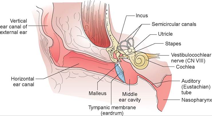
FIGURE 16.1: The external, middle, and internal ear of a dog or cat.
Copyright 2010 Cengage Learning. All Rights Reserved. May not be copied, scanned, or duplicated, in whole or in part. Due to electronic rights, some third party content may be suppressed from the eBook and/or eChapter(s).
Editorial review has deemed that any suppressed content does not materially affect the overall learning experience. Cengage Learning reserves the right to remove additional content at any time if subsequent rights restrictions require it.
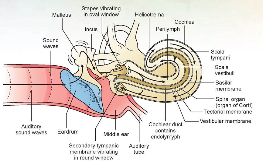
FIGURE 16.2: The ear of a dog, showing ossicles and a longitudinal cross section of the cochlea. connected to and covers the oval window of the cochlea. An acronym to help remember these names— malleus, incus, stapes—is MIS (so you will not “miss” it on a test). The ossicles act as a system of levers that transmits sound vibrations to the cochlea. In the process, the ossicles decrease the amplitude (size) of the vibrations but increase their force. As a result, the cochlea is not damaged. A tiny muscle attached to the malleus, the tensor tympani, acts to dampen vibrations. The stapedius, another muscle, acts to restrict movement of the stapes caused by loud sounds. Thus, it also prevents cochlear damage.
3. The inner ear has components that contribute to both hearing and balance. The hearing component is housed in the cochlea, a snail-shell-shaped spiral cavity within the temporal bone (Figure 16.2). Inside of this is the soft, multilayered, fluid-filled organ of Corti, which contains the receptor cells for hearing (Figure 16.3).
This organ runs the length of the cochlea in a tube called the cochlear duct, which is filled with a liquid called endolymph. On either side of this duct is a tube containing perilymph; these ducts communicate at the tip of the cochlea.The cochlear duct starts at the oval window, which is attached to the stapes, goes to the tip of the cochlea, and returns to the round window. Nothing is attached to the round window. The organ of Corti runs the length of the cochlear duct, resting on a membrane called the basilar membrane (Figure 16.4).The organ’s functional parts are the hair cells, supporting cells, and tectorial membrane. The hair cells are receptor cells, with tiny hair-like projections on their surfaces. The tectorial membrane is gelatin-like and lies gently on the hairs. The supporting cells provide physical support to the hair cells.
Sound waves transmitted to the cochlea’s oval window via the stapes cause the membrane of the window to move back and forth. This causes the perilymph around the cochlear duct to vibrate as well. The round window acts as a pressure-relief mechanism by alternately bulging out and in with the vibrations of the perilymph. The perilymph’s vibrations cause the tectorial membrane to rub against the hair cells, which bends the sensory hairs and generates nerve impulses that travel to the brain and are interpreted as sound.
The inner ear also contains the semicircular canals and vestibule. These structures are important in maintaining balance and equilibrium. The vestibule is a part of the inner ear located between the cochlea and semicircular canals. It contains two sac-like spaces called the utricle and saccule (see Figure 16.4), which are continuous with the cochlear duct and are filled with endolymph. These two sacs are surrounded by perilymph. Each sac contains an area of sensory epithelium called the macula. Each also contains hair cells and supporting cells covered by a gel-like matrix containing tiny crystals of calcium carbonate called otoliths (Figure 16.5).
Gravity causes the otoliths and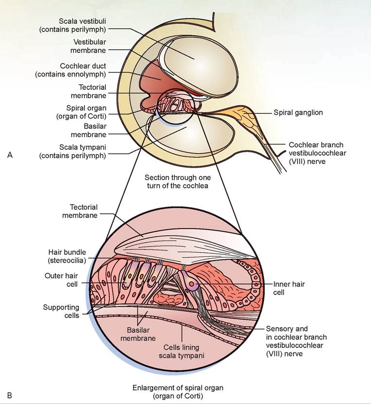
FIGURE 16.3: Cross section of the spiral organ (organ of Corti) of the cochlea of a dog. A. Section through one turn of the cochlea. B. Spiral organ, or organ of Corti.
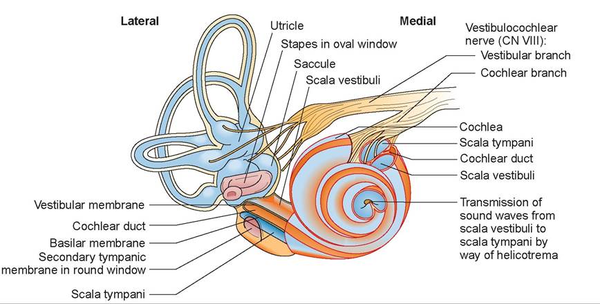
FIGURE 16.4: The inner ear of a dog with cochlea, semicircular canals, and nerve branches.
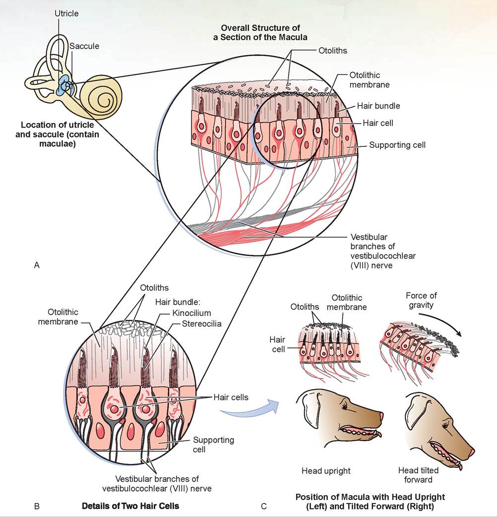
FIGURE 16.5: Receptors of the utricle and saccule of a dog and their mechanism of action. A. Section of the macula. B. Two hair cells. C. Position of macula with head upright and tilted forward.
the gelatinous matrix to put constant pressure on the hairs, so long as the head stays still. Movement of the head bends these sensory hairs, which generates nerve impulses that give the brain information about the position of the head.
Three semicircular canals, each oriented in a different plane at right angles to one another, are also filled with endolymph and surrounded by perilymph. Near the utricle end of each semicircular canal is an enlargement known as the ampulla. It contains the receptors, known as the crista ampullaris (crista). The ampulla functions in the same way as the macula of the vestibule. It consists of a cone-shaped area of hair cells and their supporting cells, which project into a gelatinous structure called the cupula (Figure 16.6). But the ampulla has no otoliths. The cupula functions as a float that moves with the endolymph as the head changes position. As the head moves in one of the planes of the semicircular canals, the inertia created causes the endolymph to lag behind the movement of the canal. The relative movement of the endolymph pulls on the cupula, bending the hairs, and generating a nerve impulse that is sent up the vestibular portion of the vestibulocochlear nerve to the brain.
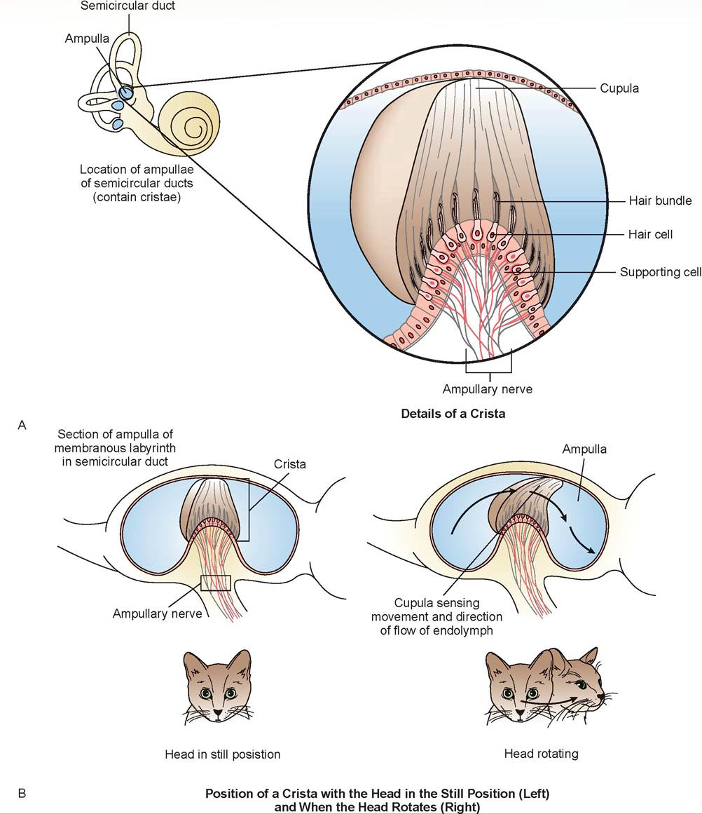
FIGURE 16.6: Receptors of the semicircular canal of a cat and their mechanism of action. A. A crista. B. Position of a crista when head is still and when it rotates.
The Eye and the Mechanisms of Vision
EXERCISE 16.2 ANATOMY AND PHYSIOLOGY OF THE EYE
Vision is a process in which light waves of differing wavelengths enter the eye and are focused on the back of the eyeball, which is lined by the retina. The retina’s nerve cells pick up the visual image, which is transmitted via the optic nerve, which crosses to the opposite side of the brain at the optic chiasm, and is then relayed through the rostral colliculi to the cortex of the occipital part of the cerebrum. This information is processed and interpreted as a picture by the brain. The image that reaches the back of the eye is upside down, but the brain converts it so the world is right-side up.
Dissection of the Sheep Eye
1. Obtain a sheep’s eye and note the fat surrounding the surface of the eye. This cushions the eye from shock in its bony orbit.
2. Identify the sclera: the tough, fibrous, external, white coat.
3. The conjunctiva is a thin, transparent membrane that covers the front portion of the sclera and lines the inside of the eyelids. It is composed of two parts: the bulbar conjunctiva, attached to the sclera, and the palpebral conjunctiva on the inner eyelids. The space between these two parts is the conjunctival sac, and they meet at the fornix.
4. The palpebrae are the eyelids. The dorsal and ventral palpebrae join at the medial canthus, or inner corner of the eye, and at the lateral canthus, or outer corner of the eye. Along the margins of the eyelids are small pores that are the openings to the tarsal glands (Meibomian glands). These glands produce a waxy substance that helps prevent tears from overflowing onto the face. The eyelashes (cilia) are more prominent on the upper lid. Domestic animals also have a third eyelid, or nictitating membrane, which is located medially between the globe of the eye and the eyelids (Figure 16.7). It is supported by an internal T-shaped piece of cartilage and is covered by conjunctiva. Behind the third eyelid, on its ocular surface, are lymph tissue and the gland of the third eyelid, an accessory lacrimal gland (tear-producing gland).
5. The lacrimal apparatus is the tear drainage route for the eye (Figure 16.8). At the medial canthus of both eyes, there are two small holes or puncta (singular: punctum), each is located a few millimeters dorsally and ventrallyjust medial to the margin of the eyelid. The dorsal punctum connects to the superior canaliculus, and the ventral punctum connects to the inferior canaliculus; these canaliculi join to form the lacrimal sac, which drains into the nasolacrimal duct. This duct exits to the nasal cavity just inside external nares on the medial surface of the wall. Patency of these ducts can be tested by placing fluorescein dye in the eyes and checking for its appearance around the nose. It is sometimes necessary to use a Wood’s light (an ultraviolet light) to observe this.
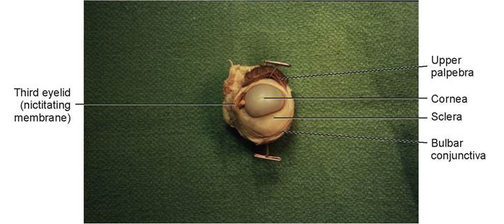
FIGURE 16.7: External view of a sheep's eye.
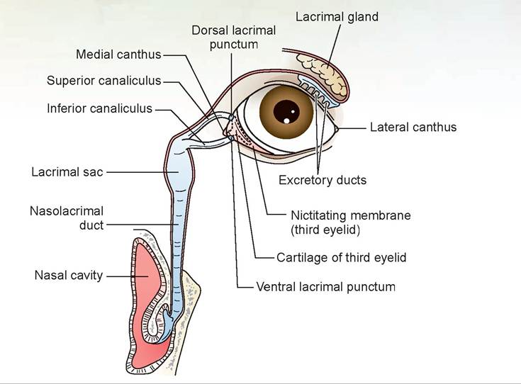
FIGURE 16.8: The eye and an internal view of the nasolacrimal system of a dog.
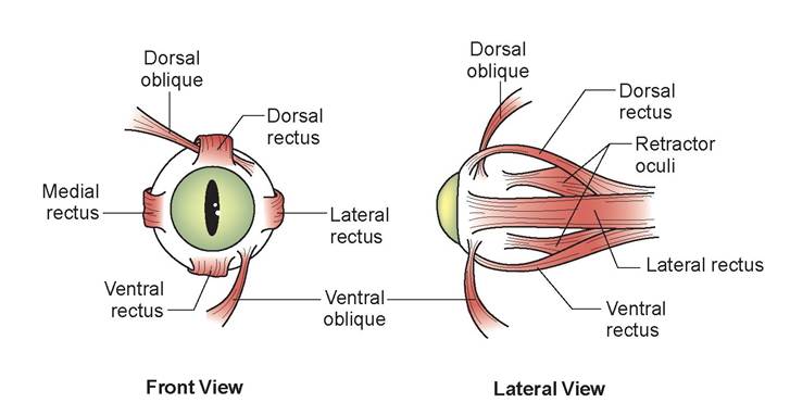
FIGURE 16.9: The extrinsic eye muscles of a dog.
6. On your specimen, the eye muscles may be attached to the globe (eyeball), but they will probably be too damaged to distinguish which muscle is which.The extraocular eye muscles (Figure 16.9) consist of four rectus muscles (the ventral, dorsal, lateral, and medial rectus muscles) and two oblique muscles (the ventral and dorsal oblique muscles). Many animals also have a retractor bulbi muscle, which humans do not. The levator palpebrae superioris muscle lifts up the upper eyelid.
7. The cornea is the anterior, transparent tissue (though it may appear opaque in your specimen because of the preservative) that attaches to the sclera at the limbus. The cornea has multiple layers. The outer epithelium is composed of stratified squamous cells that are continuous peripherally with the conjunctiva. The outer epithelium is thicker in the middle than at the periphery and shows great powers of regeneration. Beneath the epithelium is the basement membrane. The substantia propria, or stroma, is the thickest layer of the cornea and is made of transparent collagen fibrils without blood vessels. Its transparency is dependent on the exact amount of water it contains. If there is too much (corneal edema), it becomes cloudy. It contains many pain receptors, making it extremely sensitive. Below this, just deep to the stroma, is Descemefs membrane. Deep to this is the endothelium, composed of a thin layerof squamous epithelial cells (Figure 16.10).
8. The optic nerve is located on the caudal surface of the eye. This nerve has a solid, white core and is approximately 3 mm thick.
9. Make a stab incision through the sclera about 1/2 cm from the edge of the cornea. Using scissors, cut completely around the eye, parallel to the cornea.
10. You should now see the vitreous humor (vitreous body) in the caudal part of the eye, and the lens sitting in the vitreous chamber (Figure 16.11).
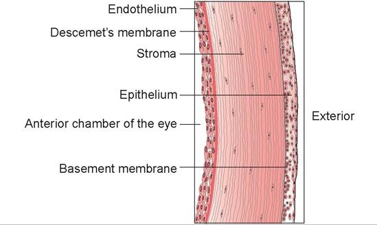
FIGURE 16.10: Layers of the cornea of a dog.
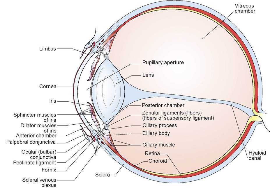
FIGURE 16.11: Sagittal section of a sheep's eye, showing vitreous humor, lens, and ciliary body.
11. Examine the interior of the rostral part of the eye. Look at the black structure around and behind the lens. This structure is the ciliary body consisting of the ciliary muscle and the ciliary processes; the latter appear as radial folds (Figures 16.11 to 16.13). Locate the suspensory ligaments, the delicate fibers connecting the ciliary body to the lens. They hold the lens in position.
12. Detach the lens from the ciliary body and remove the lens. The remnants of the suspensory ligaments can be seen attached to the lens (see Figure 16.13).
13. The iris is now visible anterior to the former position of the lens (see Figure 16.13). It also appears black (as did the ciliary body) on the caudal surface, but it may be brownish-tan on the cranial surface. Try to distinguish between the circular and radial fibers that make up the iris. These are the muscles that control the aperture opening of the iris, known as the pupil.
14. Find the space between the ciliary body and the iris; then find the space between the iris and the cornea. These are the posterior and anterior chambers, respectively. The ciliary processes produce the aqueous humor, which flows between the ciliary body and the iris, in front of the lens, and through the pupil into the anterior chamber. The aqueous humor flows out of the anterior chamber at the angle formed between the iris and cornea (the iridocorneal angle) through tiny holes in the pectinate ligament (called the spaces of Fontana), into vessels leading to the scleral venous plexus (called the canals of Schlemm), and is then absorbed back into the bloodstream. The pectinate ligament is a network of fine trabeculae connecting the iris to the inner wall of the sclera.
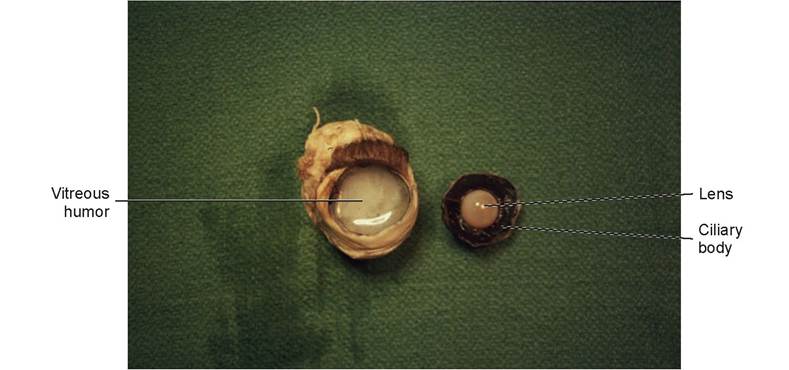
FIGURE 16.12: A transverse section of the eye of a dog.
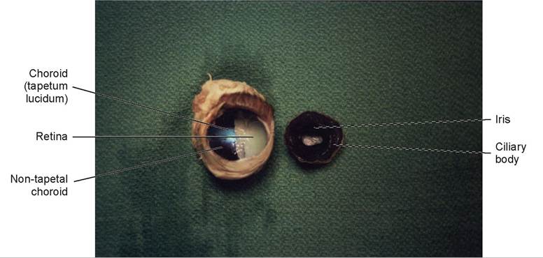
FIGURE 16.13: Sheep's eye, showing retina, choroid, and iris.
15. This first thing to note in the caudal part of the eye is the vitreous humor. During life this substance is perfectly clear (see Figure 16.13). Remove it from the eyeball.
16. The retina is the white inner coat that was covered by the vitreous humor (it is white because of the preservative). Determine the point at which the retina is attached caudally; this is the location of the optic disc, or the inner attachment of the optic nerve (see Figure 16.13).
17. The middle vascular layer of the eye is called the uvea. It consists, from anterior to posterior, of the iris, the ciliary body, and the choroid. It is located between the retina and sclera. The retina is easily separated from the choroid, the pigmented vascular layer. The choroid is continuous with the ciliary body and completely envelops the posterior hemisphere of the eyeball (the area posterior to the lens and ciliary body). It consists of six layers, which from outmost inward are the suprachoroid, the perichoroidal lymphatic space, the vascular layer, the reflective layer (tapetum lucidum), the choriocapillary layer, and the basal lamina.
The tapetum lucidum (see Figure 16.13) is a highly reflective, iridescent area. It is not present in swines or humans. The function of the tapetum lucidum is to reflect light back onto the retina. As the light passes the photoreceptors (the rods and cones) of the retina, it reflects off the tapetum and passes through the photoreceptors once again, thereby amplifying the entering light. The tapetum lucidum is also what causes an animal’s eyes to shine in the dark.
The retina is a multilayered structure that is the functional nervous tunic of the eye. It consists of five layers (Figure 16.14). Moving from the inside of the retina toward the choroid the layers are the optic nerve fibers, the ganglion cells, the bipolar cell layer, the photoreceptor layer, and the pigmented epithelial layer. The inner layer consists of axonal nerve fibers that combine to form the optic nerve at the optic disc (where all the nerve fibers converge). The disc contains only a few nerve fibers and no photoreceptors, so it is essentially a blind spot at the back of the eye.
Light must pass all the way through to the photoreceptor layer. These cells, when excited, stimulate the bipolar cells, which in turn stimulate the ganglion cells (the next layer). The axons of the ganglion cells leave the retina as a tight bundle of fibers to form the optic nerve. The photoreceptors are
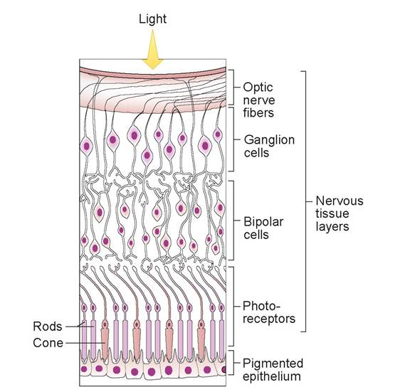
FIGURE 16.14: The layers of the retina of a cat (or other animal that has color vision). Illustration shows cones. Dogs have few cones making them functionally color-blind. Cats have enough cones to recognize colors.
neurons with dendrites modified to be sensory receptors for light. There are two types of photoreceptors, named for their shape: rods and cones. The rods are more sensitive to light and produce a somewhat coarse image in shades of gray. The cones are more sensitive to color and detail and do not function well in dim light.
Most domestic animals are described as color-blind because they have many rods and few cones. However, this description is not entirely accurate; animals can see colors to some extent, but they appear washed out or faded. They also do not perceive detail as sharply as humans. This is because humans have an area highly concentrated with cones, called the fovea centralis, in the center of the retina. Most animals need good vision to survive. The eyes of prey animals are located at the sides of their head, giving them a greater field of vision and a better chance to escape. Predators need good binocular vision to catch prey; therefore, their eyes are located more toward the front of their heads.
EXERCISE 16.3 COLOR VISION IN DOGS
This is an entertaining experiment that illustrates color vision (or the lack of it) and visual acuity in dogs.
Procedure
1. Obtain a dog that will retrieve. Place on the ground four colored balls—one each of red, green, fluorescent orange, or white—and get the dog to retrieve each of them. Then, wash each of the balls to remove any contaminating odors.
2. Blindfold the dog. In a field of green grass, place the colored balls at the 12, 3, 6, and 9 o’clock positions at the perimeter of a circle 60 ft in diameter, one ball at each position. (Note that one ball must be the same color as the grass.) Place the blindfolded dog in the center of the circle.
3. Remove the blindfold and release the dog. Observe which ball it retrieves first.
4. Remove that ball from the game and continue with the other three. Continue until all the balls are retrieved.
5. Place the white ball on a sheet of white paper, and each colored ball on sheets of colored paper of the same color. Repeat the experiment. This should be a test of visual acuity.
Questions
1. Which ball did the dog retrieve first?
2. Why would placing the balls on sheets of paper of the same color be a test of visual acuity?
Discussion
The ball the dog should see easiest is the white ball. Even the fluorescent orange ball will have a grayscale appearance similar to that of the green and red balls, and to that of the grass. It is easy for a human to see, but not a dog. Fluorescent-orange boat bumpers are great to teach a retriever hand signals because you can see the bumper from a distance in high grass, but the dog cannot and needs your help to find it. Placing the balls on similarly colored sheets of paper removes the contrasting background, and the dog must then rely on its sense of visual acuity—that is, how well it sees the shadow cast around the ball—to find it.
Clinical Significance
The amount of aqueous humor in the anterior chamber of the eye determines the eye’s intraocular pressure. If the animal does not have the ability to absorb this liquid, the volume within the eye will increase, and so will the pressure. This pressure causes a disease condition called glaucoma. It results in degenerative changes in the retina and optic nerve and eventually leads to blindness.
When a veterinary ophthalmologist examines an animal’s eye, one of the tests performed is to visualize the angle between the cornea and iris (the iridocorneal angle). This is done using a slit lamp.
A narrow beam of light is passed through the cornea and viewed from an angle perpendicular to the beam. Often glaucoma is caused by a narrowed angle between the iris and cornea, which impairs the drainage through the spaces of Fontana and into the canals of Schlemm. We can measure the pressure within the ocular chamber using a tonometer. Pressures greater than 25 to 30 mmHg in dogs, or more than 30 mmHg in cats, indicate glaucoma. Increased pressures may also be secondary to other diseases, such as primary lens luxation, anterior uveitis, and hyphema.
Most forms of glaucoma are best treated surgically. Surgery can enhance the outflow or reduce the production of aqueous humor. Unfortunately, veterinarians often are presented with cases that are already quite advanced, and treatment is unsuccessful.

oc, Tom Foley is on his way in with his Rottweiler, Granite. He's just been hit by a train,”
my receptionist rushed in to tell me. “He'll be here in about 30 minutes,” she said.
k-? ' “You mean a real train? Uh... like a real choo-choo?” I asked. I have always liked using the most technical terms with my staff; it makes us appear professional.
“Yep.”
“How bad is it?” I asked.
“Don't know; he didn't say. He just hung up in a hurry,” she told me.
Oh no! I thought. The last time I had one of those, the dog had chest trauma, was throwing all sorts of abnormal electrocardiogram wave patterns, and was hemorrhaging internally. I rushed into the treatment room to tell my technician what was going on and to get the trauma cart ready. “This could be a bad one,” I told her. I turned up the radio when we heard the news flash come in.
“We advise you to route around Highway 99 West at Glen Market Road. We have reports that the northbound Union Pacific express is stalled at this intersection. It seems an animal was injured and is on its way to an emergency veterinary hospital. We will keep you posted on these developments as they come in. This is Sharon Schilling, live on the scene for KPTX.”
I had thought I was going to get to go home early this evening. It had been a relatively slow day; a few vaccinations, 14 cases in a row of flea allergic dermatitis, and a man wanting codeine for his non-coughing dog with kennel cough.
“He's here, Doctor,” the receptionist told me.
We rushed out to help. Granite was standing in the waiting room, tongue hanging out the side of his mouth, panting and drooling on the floor, and wagging his tail! He jumped up, as he usually does, putting his front feet on the counter top to look over at the receptionist. His left eye had popped out of its socket, a condition called proptosis. Tom had had the good sense to hobble Granite by tying his front feet loosely together, and he had covered the eye with a moist cloth to protect it. He had completed a first aid course at work and was using his training on his dog. Smart man!
“I heard the report on the radio about a train on 99, is that the same one?” I asked him.
“Yeah, he ran out in front of it, got hit on the back of the head and knocked about 20 feet.” That explained the dog. What about the train? I thought.
“I don't know why it stopped,” Tom added. “Maybe he derailed it.” He laughed.
We checked Granite over and found nothing else wrong. We replaced his eye and sewed the lids together to protect it and hold it in place. I injected some aqueous cortisone behind the eye and explained to Tom that it would take a while for the hemorrhaging and swelling in the fat pad behind Granite's eye to go down. Because we repaired the eye quickly, he would probably not lose his sight, which he didn't. The funny thing about Granite was, he didn't even act like he had a headache.
We found out later that the engineer of the train saw the dog running toward the track, then tried to look back to see if he had hit it. In the process, he banged his head and cut a huge hole in his scalp. He probably had a worse headache than Granite. But then, maybe his head wasn't as hard!
Summary
The focus of this chapter was the senses of sight, balance and equilibrium, and hearing. The special senses of smell and taste were covered briefly in previous chapters. You learned the anatomy of the external, middle, and internal ears using the provided drawings and a plastic model. The physiological mechanism of hearing and the transmission of nerve impulses to the brain also were discussed. Using the illustrations provided, the mechanisms by which the semicircular canals and the vestibule in the inner ear recognize changes in posture and spatial orientation were covered. You were able to dissect a sheep's eye and learn its anatomical structures. Illustrations were used to describe how the retina works to facilitate vision. Discussion also included the mechanism of aqueous humor production and resorption, the microscopic anatomy of the cornea, and the mechanism of sight. Finally, a fun exercise using a live animal was utilized to illustrate the lack of color vision and the extent of visual acuity in dogs. In this you learned that dogs have trouble seeing items of similar gray-scale values, like green grass and orange balls, but that objects having substantial black-and-white contrast are much easier for them to see.
REVIEW QUESTIONS
1. Sound waves hitting the eardrum are translated into sensory impulses that are transmitted through what structure to the temporal part of the brain?
2. Name the three anatomical and functional parts of the ear.
3. What is the name of the ear flap?
4. Give both names of the structure that divides the horizontal ear canal from the middle ear.
5. What structure connects the middle ear to the nasopharynx, and why is its function necessary?
6. Name the three bones of the middle ear in order, from exterior to interior.
7. Name the two organs of the inner ear that are responsible for balance and equilibrium.
8. What are the little stones in the inner ear that stimulate the hair cells called?
9. How are the semicircular canals arranged, and how is that arrangement of significance in their function?
10. What is the name of the bony location of the eye?
11. What is the fibrous outer coat of the eye called?
12. Differentiate between the bulbar conjunctiva and the palpebral conjunctiva. What is the site where these two structures meet called?
13. What are the lateral and medial corners of the eyes called?
14. Give both names of the glands that have small pores on the margins of the eyelid.
15. What is the other name for the third eyelid?
16. Name the apparatus (and its component structures) that drains tears from the eyelids to the nose.
17. Name the layers of the cornea.
18. What is the name of the space between the cornea and the pupil?
19. What is the name of the space between the inner iris and the ciliary body?
20. Where is the vitreous humor located?
21. Name the innermost nerve cell layer at the back of the globe of the eye. The white dot at the center of this layer is called what? This white dot connects to what structure?
22. What is the choroid layer?
23. What is the name of the shiny, reflective layer of the choroid, and what is its function?
24. Name the two types of photoreceptors, and indicate which type is responsible for black and white vision, and which type is responsible for seeing color.
25. What anatomical defect is responsible for the disease called glaucoma?